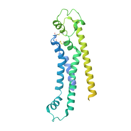
6LMW | pdb_00006lmw

Cryo-EM structure of the CALHM chimeric construct (8-mer)


Experimental Data Snapshot

  • Method: ELECTRON MICROSCOPY
  • Resolution: 3.40 Å
  • Aggregation State: PARTICLE 
  • Reconstruction Method: SINGLE PARTICLE 

wwPDB Validation 3D Report Full Report

Validation slider image for 6LMW

This is version 1.3 of the entry. See complete history. 

Literature

Cryo-EM structures of calcium homeostasis modulator channels in diverse oligomeric assemblies.

Demura, K., Kusakizako, T., Shihoya, W., Hiraizumi, M., Nomura, K., Shimada, H., Yamashita, K., Nishizawa, T., Taruno, A., Nureki, O.

(2020) Sci Adv 6: eaba8105-eaba8105

  • DOI: https://doi.org/10.1126/sciadv.aba8105
  • Primary Citation Related Structures: 
    6LMT, 6LMU, 6LMV, 6LMW, 6LMX

  • PubMed Abstract: 

    Calcium homeostasis modulator (CALHM) family proteins are Ca 2+ -regulated adenosine triphosphate (ATP)-release channels involved in neural functions including neurotransmission in gustation. Here, we present the cryo-electron microscopy (EM) structures of killifish CALHM1, human CALHM2, and Caenorhabditis elegans CLHM-1 at resolutions of 2.66, 3.4, and 3.6 Å, respectively. The CALHM1 octamer structure reveals that the N-terminal helix forms the constriction site at the channel pore in the open state and modulates the ATP conductance. The CALHM2 undecamer and CLHM-1 nonamer structures show the different oligomeric stoichiometries among CALHM homologs. We further report the cryo-EM structures of the chimeric construct, revealing that the intersubunit interactions at the transmembrane domain (TMD) and the TMD-intracellular domain linker define the oligomeric stoichiometry. These findings advance our understanding of the ATP conduction and oligomerization mechanisms of CALHM channels.


  • Organizational Affiliation: 
    • Department of Biological Sciences, Graduate School of Science, The University of Tokyo, 7-3-1 Hongo, Bunkyo-ku, Tokyo 113-0033, Japan.

Macromolecule Content 

  • Total Structure Weight: 299.34 kDa 
  • Atom Count: 11,624 
  • Modeled Residue Count: 1,472 
  • Deposited Residue Count: 2,608 
  • Unique protein chains: 1

Macromolecules

Find similar proteins by:|  3D Structure
Entity ID: 1
MoleculeChains  Sequence LengthOrganismDetailsImage
Calcium homeostasis modulator 1,Calcium homeostasis modulator protein 2
A, B, C, D, E
A, B, C, D, E, F, G, H
326Oryzias latipes, Homo sapiens
This entity is chimeric
Mutation(s): 0 
Gene Names: CALHM1, CALHM2, FAM26B
Membrane Entity: Yes 
UniProt & NIH Common Fund Data Resources
Find proteins for H2MCM1 (Oryzias latipes)
Explore H2MCM1 
Go to UniProtKB:  H2MCM1
Find proteins for Q9HA72 (Homo sapiens)
Explore Q9HA72 
Go to UniProtKB:  Q9HA72
PHAROS:  Q9HA72
GTEx:  ENSG00000138172 
Entity Groups
Sequence Clusters30% Identity50% Identity70% Identity90% Identity95% Identity100% Identity
UniProt GroupsH2MCM1Q9HA72
Sequence Annotations
Expand
Reference Sequence

Experimental Data & Validation

Experimental Data

  • Method: ELECTRON MICROSCOPY
  • Resolution: 3.40 Å
  • Aggregation State: PARTICLE 
  • Reconstruction Method: SINGLE PARTICLE 
EM Software:
TaskSoftware PackageVersion
MODEL REFINEMENTPHENIX

Structure Validation

View Full Validation Report


Entry History 

Deposition Data

Revision History  (Full details and data files)

  • Version 1.0: 2020-07-29
    Type: Initial release
  • Version 1.1: 2020-09-09
    Changes: Database references
  • Version 1.2: 2024-11-20
    Changes: Data collection, Database references, Structure summary
  • Version 1.3: 2025-07-02
    Changes: Data collection