6LAX | pdb_00006lax

the mutant SAM-VI riboswitch (U6C) bound to SAM


Experimental Data Snapshot

  • Method: X-RAY DIFFRACTION
  • Resolution: 2.70 Å
  • R-Value Free: 
    0.239 (Depositor), 0.241 (DCC) 
  • R-Value Work: 
    0.189 (Depositor), 0.194 (DCC) 
  • R-Value Observed: 
    0.191 (Depositor) 

Starting Model: experimental
View more details

wwPDB Validation 3D Report Full Report

Validation slider image for 6LAX

Ligand Structure Quality Assessment 


This is version 1.3 of the entry. See complete history

Literature

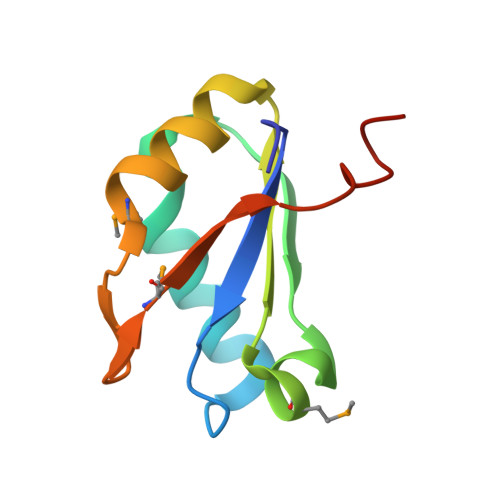
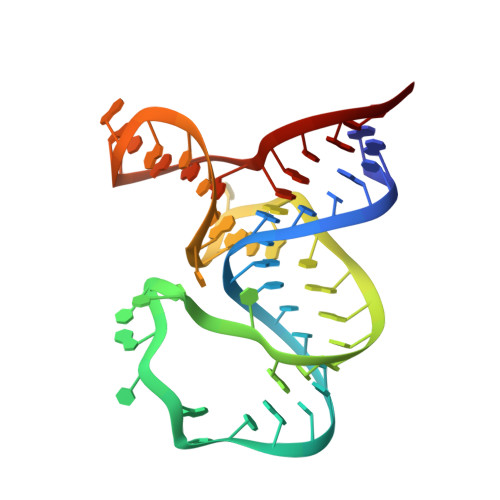
SAM-VI riboswitch structure and signature for ligand discrimination.

Sun, A.Gasser, C.Li, F.Chen, H.Mair, S.Krasheninina, O.Micura, R.Ren, A.

(2019) Nat Commun 10: 5728-5728

  • DOI: https://doi.org/10.1038/s41467-019-13600-9
  • Primary Citation Related Structures: 
    6LAS, 6LAU, 6LAX, 6LAZ

  • PubMed Abstract: 

    Riboswitches are metabolite-sensing, conserved domains located in non-coding regions of mRNA that are central to regulation of gene expression. Here we report the first three-dimensional structure of the recently discovered S-adenosyl-L-methionine responsive SAM-VI riboswitch. SAM-VI adopts a unique fold and ligand pocket that are distinct from all other known SAM riboswitch classes. The ligand binds to the junctional region with its adenine tightly intercalated and Hoogsteen base-paired. Furthermore, we reveal the ligand discrimination mode of SAM-VI by additional X-ray structures of this riboswitch bound to S-adenosyl-L-homocysteine and a synthetic ligand mimic, in combination with isothermal titration calorimetry and fluorescence spectroscopy to explore binding thermodynamics and kinetics. The structure is further evaluated by analysis of ligand binding to SAM-VI mutants. It thus provides a thorough basis for developing synthetic SAM cofactors for applications in chemical and synthetic RNA biology.


  • Organizational Affiliation
    • Life Sciences Institute, Zhejiang University, 310058, Hangzhou, Zhejiang, China.

Macromolecule Content 

  • Total Structure Weight: 69.22 kDa 
  • Atom Count: 4,661 
  • Modeled Residue Count: 385 
  • Deposited Residue Count: 389 
  • Unique protein chains: 1
  • Unique nucleic acid chains: 1

Macromolecules


Find similar proteins by:|  3D Structure
Entity ID: 2
MoleculeChains  Sequence LengthOrganismDetailsImage
U1 small nuclear ribonucleoprotein AB [auth D],
C,
D [auth E]
93Homo sapiensMutation(s): 3 
Gene Names: SNRPA
UniProt & NIH Common Fund Data Resources
Find proteins for P09012 (Homo sapiens)
Explore P09012 
Go to UniProtKB:  P09012
PHAROS:  P09012
GTEx:  ENSG00000077312 
Entity Groups
Sequence Clusters30% Identity50% Identity70% Identity90% Identity95% Identity100% Identity
UniProt GroupP09012
Sequence Annotations
Expand
Reference Sequence
Find similar nucleic acids by:  (by identity cutoff) 
Entity ID: 1
MoleculeChains LengthOrganismImage
RNA (55-MER)A,
E [auth B]
55Bifidobacterium angulatum
Sequence Annotations
Expand
Reference Sequence

Small Molecules

Modified Residues  1 Unique
IDChains TypeFormula2D DiagramParent
MSE
Query on MSE
B [auth D],
C,
D [auth E]
L-PEPTIDE LINKINGC5 H11 N O2 SeMET

Experimental Data & Validation

Experimental Data

  • Method: X-RAY DIFFRACTION
  • Resolution: 2.70 Å
  • R-Value Free:  0.239 (Depositor), 0.241 (DCC) 
  • R-Value Work:  0.189 (Depositor), 0.194 (DCC) 
  • R-Value Observed: 0.191 (Depositor) 
Space Group: P 1 21 1
Unit Cell:
Length ( Å )Angle ( ˚ )
a = 45.892α = 90
b = 86.942β = 99.19
c = 93.626γ = 90
Software Package:
Software NamePurpose
HKL-2000data scaling
PHENIXrefinement
PDB_EXTRACTdata extraction
HKL-2000data reduction
PHASERphasing

Structure Validation

View Full Validation Report


Ligand Structure Quality Assessment 


Entry History 

& Funding Information

Deposition Data


Funding OrganizationLocationGrant Number
National Natural Science Foundation of ChinaChina31870810, 91640104 and 31670826

Revision History  (Full details and data files)

  • Version 1.0: 2020-01-01
    Type: Initial release
  • Version 1.1: 2023-11-22
    Changes: Data collection, Database references, Refinement description
  • Version 1.2: 2024-10-23
    Changes: Structure summary
  • Version 1.3: 2025-03-12
    Changes: Database references