6L4G | pdb_00006l4g

Crystal structure of human NDRG3 I171M/S176H mutant


Experimental Data Snapshot

  • Method: X-RAY DIFFRACTION
  • Resolution: 3.30 Å
  • R-Value Free: 
    0.226 (Depositor), 0.214 (DCC) 
  • R-Value Work: 
    0.196 (Depositor), 0.176 (DCC) 

Starting Model: experimental
View more details

wwPDB Validation 3D Report Full Report

Validation slider image for 6L4G

This is version 1.3 of the entry. See complete history

Literature

Structural and Biophysical Analyses of Human N-Myc Downstream-Regulated Gene 3 (NDRG3) Protein.

Kim, K.R.Kim, K.A.Park, J.S.Jang, J.Y.Choi, Y.Lee, H.H.Lee, D.C.Park, K.C.Yeom, Y.I.Kim, H.J.Han, B.W.

(2020) Biomolecules 10

  • DOI: https://doi.org/10.3390/biom10010090
  • Primary Citation Related Structures: 
    6L4B, 6L4G, 6L4H

  • PubMed Abstract: 

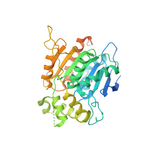
    The N-Myc downstream-regulated gene (NDRG) family belongs to the α/β-hydrolase fold and is known to exert various physiologic functions in cell proliferation, differentiation, and hypoxia-induced cancer metabolism. In particular, NDRG3 is closely related to proliferation and migration of prostate cancer cells, and recent studies reported its implication in lactate-triggered hypoxia responses or tumorigenesis. However, the underlying mechanism for the functions of NDRG3 remains unclear. Here, we report the crystal structure of human NDRG3 at 2.2 Å resolution, with six molecules in an asymmetric unit. While NDRG3 adopts the α/β-hydrolase fold, complete substitution of the canonical catalytic triad residues to non-reactive residues and steric hindrance around the pseudo-active site seem to disable the α/β-hydrolase activity. While NDRG3 shares a high similarity to NDRG2 in terms of amino acid sequence and structure, NDRG3 exhibited remarkable structural differences in a flexible loop corresponding to helix α6 of NDRG2 that is responsible for tumor suppression. Thus, this flexible loop region seems to play a distinct role in oncogenic progression induced by NDRG3. Collectively, our studies could provide structural and biophysical insights into the molecular characteristics of NDRG3.


  • Organizational Affiliation
    • Research Institute of Pharmaceutical Sciences, College of Pharmacy, Seoul National University, Seoul 08826, Korea.

Macromolecule Content 

  • Total Structure Weight: 83.04 kDa 
  • Atom Count: 4,350 
  • Modeled Residue Count: 560 
  • Deposited Residue Count: 750 
  • Unique protein chains: 1

Macromolecules

Find similar proteins by:|  3D Structure
Entity ID: 1
MoleculeChains  Sequence LengthOrganismDetailsImage
Protein NDRG3
A, B
375Homo sapiensMutation(s): 2 
Gene Names: NDRG3
UniProt & NIH Common Fund Data Resources
Find proteins for Q9UGV2 (Homo sapiens)
Explore Q9UGV2 
Go to UniProtKB:  Q9UGV2
PHAROS:  Q9UGV2
GTEx:  ENSG00000101079 
Entity Groups
Sequence Clusters30% Identity50% Identity70% Identity90% Identity95% Identity100% Identity
UniProt GroupQ9UGV2
Sequence Annotations
Expand
Reference Sequence

Experimental Data & Validation

Experimental Data

  • Method: X-RAY DIFFRACTION
  • Resolution: 3.30 Å
  • R-Value Free:  0.226 (Depositor), 0.214 (DCC) 
  • R-Value Work:  0.196 (Depositor), 0.176 (DCC) 
Space Group: P 32 2 1
Unit Cell:
Length ( Å )Angle ( ˚ )
a = 100.387α = 90
b = 100.387β = 90
c = 111.756γ = 120
Software Package:
Software NamePurpose
REFMACrefinement
HKL-2000data reduction
HKL-2000data scaling
PHENIXphasing

Structure Validation

View Full Validation Report


Entry History 

Deposition Data

Revision History  (Full details and data files)

  • Version 1.0: 2020-08-26
    Type: Initial release
  • Version 1.1: 2021-07-07
    Changes: Data collection
  • Version 1.2: 2023-11-22
    Changes: Data collection, Database references, Derived calculations, Refinement description
  • Version 1.3: 2024-11-13
    Changes: Structure summary