6L1N | pdb_00006l1n

Substrate bound BacF structure from Bacillus subtillis


Experimental Data Snapshot

  • Method: X-RAY DIFFRACTION
  • Resolution: 2.00 Å
  • R-Value Free: 
    0.239 (Depositor), 0.242 (DCC) 
  • R-Value Work: 
    0.182 (Depositor), 0.187 (DCC) 
  • R-Value Observed: 
    0.184 (Depositor) 

Starting Model: experimental
View more details

wwPDB Validation 3D Report Full Report

Validation slider image for 6L1N

Ligand Structure Quality Assessment 


This is version 1.1 of the entry. See complete history

Literature

Structural insights into the catalytic mechanism of Bacillus subtilis BacF.

Deshmukh, A.Gopal, B.

(2020) Acta Crystallogr F Struct Biol Commun 76: 145-151

  • DOI: https://doi.org/10.1107/S2053230X20001636
  • Primary Citation Related Structures: 
    6L1L, 6L1N, 6L1O

  • PubMed Abstract: 

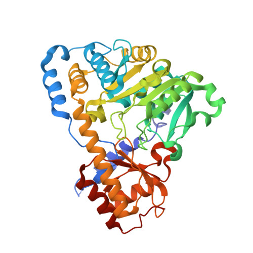
    The nonribosomal biosynthesis of the dipeptide antibiotic bacilysin is achieved by the concerted action of multiple enzymes in the Bacillus subtilis bac operon. BacF (YwfG), encoded by the bacF gene, is a fold type I pyridoxal 5-phosphate (PLP)-dependent stereospecific transaminase. Activity assays with L-phenylalanine and 4-hydroxyphenylpyruvic acid (4HPP), a chemical analogue of tetrahydrohydroxyphenylpyruvic acid (H 4 HPP), revealed stereospecific substrate preferences, a finding that was consistent with previous reports on the role of this enzyme in bacilysin synthesis. The crystal structure of this dimeric enzyme was determined in its apo form as well as in substrate-bound and product-bound conformations. Two ligand-bound structures were determined by soaking BacF crystals with substrates (L-phenylalanine and 4-hydroxyphenylpyruvate). These structures reveal multiple catalytic steps: the internal aldimine with PLP and two external aldimine conformations that show the rearrangement of the external aldimine to generate product (L-tyrosine). Together, these structural snapshots provide an insight into the catalytic mechanism of this transaminase.


  • Organizational Affiliation
    • Molecular Biophysics Unit, Indian Institute of Science, Bangalore, Karnataka 560 012, India.

Macromolecule Content 

  • Total Structure Weight: 90.15 kDa 
  • Atom Count: 6,198 
  • Modeled Residue Count: 786 
  • Deposited Residue Count: 798 
  • Unique protein chains: 1

Macromolecules

Find similar proteins by:|  3D Structure
Entity ID: 1
MoleculeChains  Sequence LengthOrganismDetailsImage
Aminotransferase
A, B
399Bacillus subtilisMutation(s): 0 
Gene Names: B4417_2089
EC: 2.6.1
UniProt
Find proteins for P39643 (Bacillus subtilis (strain 168))
Explore P39643 
Go to UniProtKB:  P39643
Entity Groups
Sequence Clusters30% Identity50% Identity70% Identity90% Identity95% Identity100% Identity
UniProt GroupP39643
Sequence Annotations
Expand
Reference Sequence

Experimental Data & Validation

Experimental Data

  • Method: X-RAY DIFFRACTION
  • Resolution: 2.00 Å
  • R-Value Free:  0.239 (Depositor), 0.242 (DCC) 
  • R-Value Work:  0.182 (Depositor), 0.187 (DCC) 
  • R-Value Observed: 0.184 (Depositor) 
Space Group: P 1 21 1
Unit Cell:
Length ( Å )Angle ( ˚ )
a = 71.29α = 90
b = 56.91β = 97.86
c = 78.5γ = 90
Software Package:
Software NamePurpose
SCALAdata scaling
REFMACrefinement
PDB_EXTRACTdata extraction
iMOSFLMdata reduction
PHASERphasing

Structure Validation

View Full Validation Report


Ligand Structure Quality Assessment 


Entry History 

& Funding Information

Deposition Data


Funding OrganizationLocationGrant Number
Department of Biotechnology (India)India--

Revision History  (Full details and data files)

  • Version 1.0: 2020-03-18
    Type: Initial release
  • Version 1.1: 2023-11-22
    Changes: Data collection, Database references, Refinement description