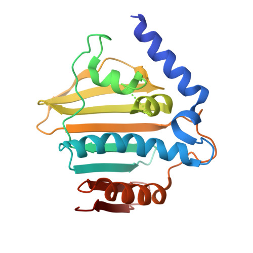
6KZV

Crystal structure of E.coli DNA gyrase B in complex with 2-oxo-1,2-dihydroquinoline derivative


Experimental Data Snapshot

  • Method: X-RAY DIFFRACTION
  • Resolution: 2.40 Å
  • R-Value Free: 0.260 
  • R-Value Work: 0.196 
  • R-Value Observed: 0.199 

wwPDB Validation   3D Report Full Report


Ligand Structure Quality Assessment 


This is version 1.2 of the entry. See complete history


Literature

Lead Identification of 8-(Methylamino)-2-oxo-1,2-dihydroquinoline Derivatives as DNA Gyrase Inhibitors: Hit-to-Lead Generation Involving Thermodynamic Evaluation.

Ushiyama, F.Amada, H.Takeuchi, T.Tanaka-Yamamoto, N.Kanazawa, H.Nakano, K.Mima, M.Masuko, A.Takata, I.Hitaka, K.Iwamoto, K.Sugiyama, H.Ohtake, N.

(2020) ACS Omega 5: 10145-10159

  • DOI: https://doi.org/10.1021/acsomega.0c00865
  • Primary Citation of Related Structures:  
    6KZV, 6KZX, 6KZZ, 6L01

  • PubMed Abstract: 

    DNA gyrase and topoisomerase IV are well-validated pharmacological targets, and quinolone antibacterial drugs are marketed as their representative inhibitors. However, in recent years, resistance to these existing drugs has become a problem, and new chemical classes of antibiotics that can combat resistant strains of bacteria are strongly needed. In this study, we applied our hit-to-lead (H2L) chemistry for the identification of a new chemical class of GyrB/ParE inhibitors by efficient use of thermodynamic parameters. Investigation of the core fragments obtained by fragmentation of high-throughput screening hit compounds and subsequent expansion of the hit fragment was performed using isothermal titration calorimetry (ITC). The 8-(methylamino)-2-oxo-1,2-dihydroquinoline derivative 13e showed potent activity against Escherichia coli DNA gyrase with an IC 50 value of 0.0017 μM. In this study, we demonstrated the use of ITC for primary fragment screening, followed by structural optimization to obtain lead compounds, which advanced into further optimization for creating novel antibacterial agents.


  • Organizational Affiliation

    Chemistry Laboratories, Taisho Pharmaceutical Company Ltd., 1-403, Yoshino-Cho, Kita-Ku, Saitama, Saitama 331-9530, Japan.


Macromolecules
Find similar proteins by:  (by identity cutoff)  |  3D Structure
Entity ID: 1
MoleculeChains Sequence LengthOrganismDetailsImage
DNA gyrase subunit B220Escherichia coliMutation(s): 0 
Gene Names: gyrBC9Z04_12785
EC: 5.6.2.2
UniProt
Find proteins for P0AES6 (Escherichia coli (strain K12))
Explore P0AES6 
Go to UniProtKB:  P0AES6
Entity Groups  
Sequence Clusters30% Identity50% Identity70% Identity90% Identity95% Identity100% Identity
UniProt GroupP0AES6
Sequence Annotations
Expand
  • Reference Sequence
Small Molecules
Ligands 1 Unique
IDChains Name / Formula / InChI Key2D Diagram3D Interactions
E0F (Subject of Investigation/LOI)
Query on E0F

Download Ideal Coordinates CCD File 
B [auth A]~{N}-[2-[[cyclohexyl(methyl)amino]methyl]phenyl]-2-oxidanylidene-1~{H}-quinoline-3-carboxamide
C24 H27 N3 O2
HXOAPOMHVOADBB-UHFFFAOYSA-N
Binding Affinity Annotations 
IDSourceBinding Affinity
E0F Binding MOAD:  6KZV IC50: 5400 (nM) from 1 assay(s)
Experimental Data & Validation

Experimental Data

  • Method: X-RAY DIFFRACTION
  • Resolution: 2.40 Å
  • R-Value Free: 0.260 
  • R-Value Work: 0.196 
  • R-Value Observed: 0.199 
  • Space Group: P 41
Unit Cell:
Length ( Å )Angle ( ˚ )
a = 53.444α = 90
b = 53.444β = 90
c = 67.884γ = 90
Software Package:
Software NamePurpose
REFMACrefinement
CrystalCleardata reduction
CrystalCleardata scaling
MOLREPphasing

Structure Validation

View Full Validation Report


Ligand Structure Quality Assessment 


Entry History 

Deposition Data

Revision History  (Full details and data files)

  • Version 1.0: 2020-05-06
    Type: Initial release
  • Version 1.1: 2020-05-27
    Changes: Database references
  • Version 1.2: 2023-11-22
    Changes: Data collection, Database references, Refinement description