6KX1 | pdb_00006kx1

Crystal structure of SN-101 mAb in complex with MUC1 glycopeptide


Experimental Data Snapshot

  • Method: X-RAY DIFFRACTION
  • Resolution: 1.77 Å
  • R-Value Free: 
    0.229 (Depositor), 0.230 (DCC) 
  • R-Value Work: 
    0.177 (Depositor), 0.179 (DCC) 
  • R-Value Observed: 
    0.179 (Depositor) 

Starting Model: experimental
View more details

wwPDB Validation 3D Report Full Report

Validation slider image for 6KX1

Ligand Structure Quality Assessment 


This is version 1.2 of the entry. See complete history. 

Literature

A straightforward approach to antibodies recognising cancer specific glycopeptidic neoepitopes

Wakui, H., Tanaka, Y., Ose, T., Matsumoto, I., Kato, K., Min, Y., Tachibana, T., Sato, M., Naruchi, K., Martin, F.G., Hinou, H., Nishimura, S.-I.

(2020) Chem Sci 11: 4999-5006

Macromolecule Content 

  • Total Structure Weight: 53.66 kDa 
  • Atom Count: 3,619 
  • Modeled Residue Count: 440 
  • Deposited Residue Count: 491 
  • Unique protein chains: 3

Macromolecules

Find similar proteins by:|  3D Structure
Entity ID: 1
MoleculeChains  Sequence LengthOrganismDetailsImage
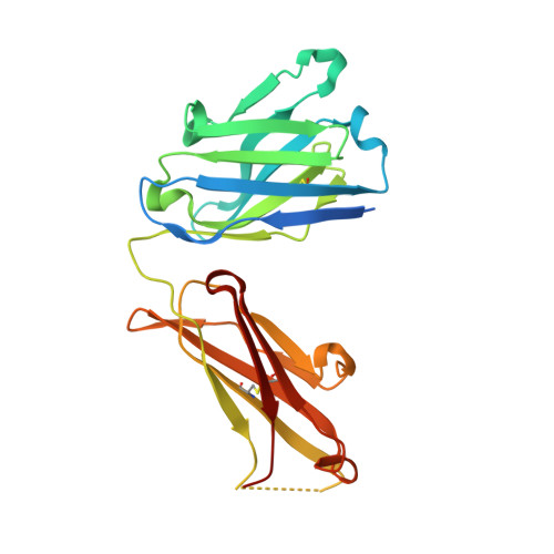
Fab Fragment-SN-101-Heavy chain236Mus musculusMutation(s): 0 
Entity Groups
Sequence Clusters30% Identity50% Identity70% Identity90% Identity95% Identity100% Identity
Sequence Annotations
Expand
Reference Sequence
Find similar proteins by:|  3D Structure
Entity ID: 2
MoleculeChains  Sequence LengthOrganismDetailsImage
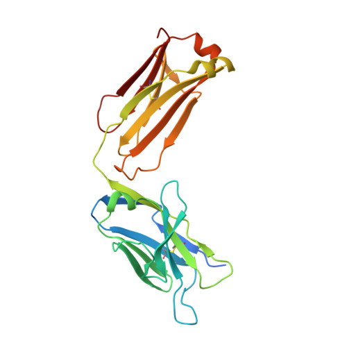
Fab Fragment-SN-101-Light chain239Mus musculusMutation(s): 0 
Entity Groups
Sequence Clusters30% Identity50% Identity70% Identity90% Identity95% Identity100% Identity
Sequence Annotations
Expand
Reference Sequence
Find similar proteins by:  Sequence   |   3D Structure  
Entity ID: 3
MoleculeChains  Sequence LengthOrganismDetailsImage
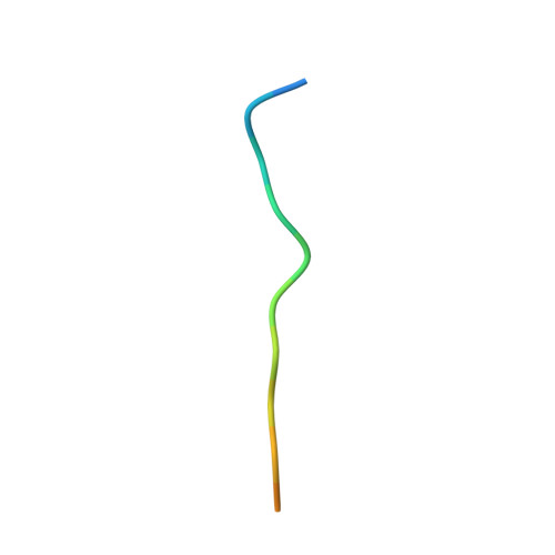
Synthetic MUC1 glycopepide16Homo sapiensMutation(s): 0 
UniProt & NIH Common Fund Data Resources
Find proteins for P15941 (Homo sapiens)
Explore P15941 
Go to UniProtKB:  P15941
PHAROS:  P15941
GTEx:  ENSG00000185499 
Entity Groups
Sequence Clusters30% Identity50% Identity70% Identity90% Identity95% Identity100% Identity
UniProt GroupP15941
Glycosylation
Glycosylation Sites: 1Go to GlyGen: P15941-1
Sequence Annotations
Expand
Reference Sequence

Small Molecules

Ligands 1 Unique
IDChains Name / Formula / InChI Key2D Diagram3D Interactions
A2G
(Subject of Investigation/LOI)

Query on A2G


Download:Ideal Coordinates CCD File
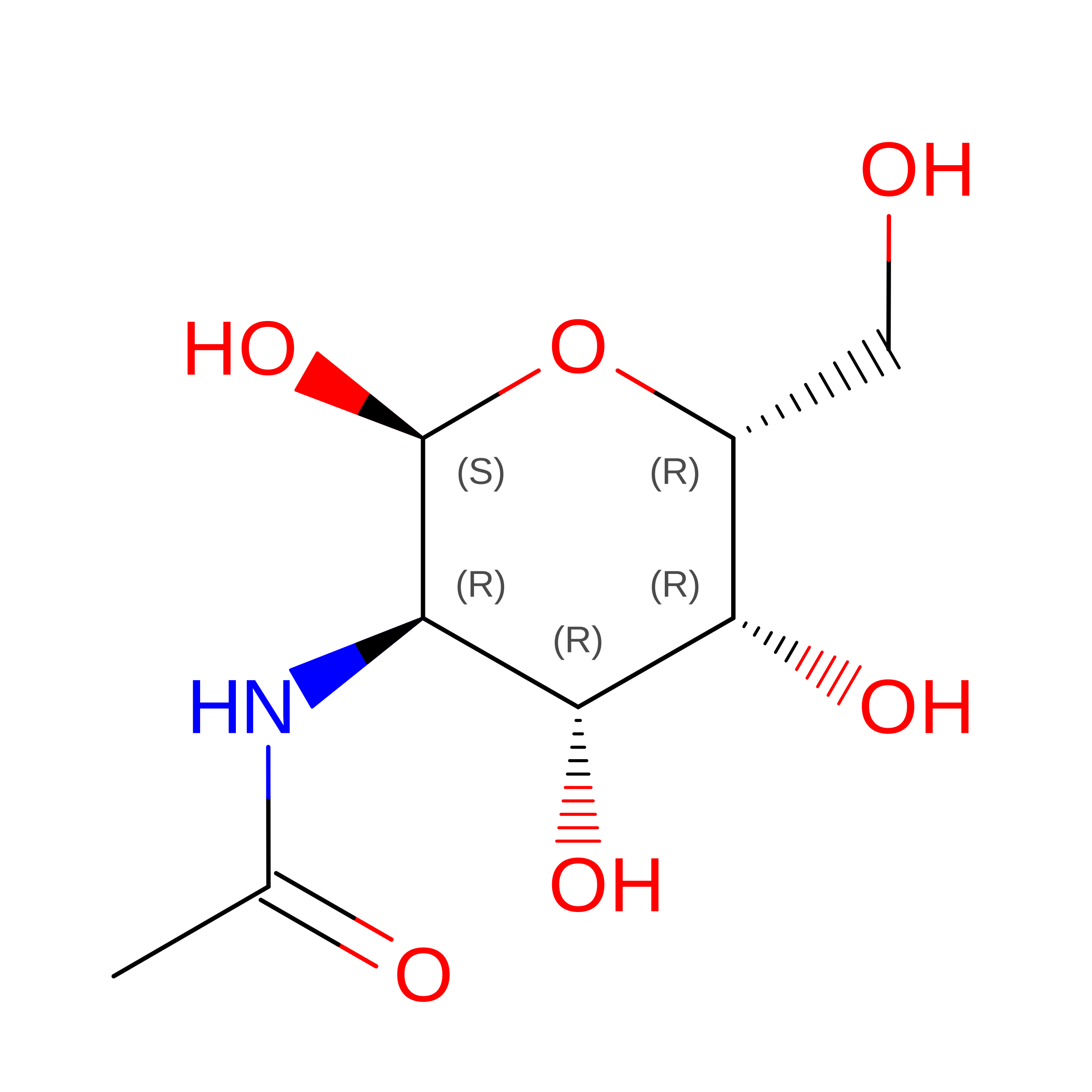
D [auth C]2-acetamido-2-deoxy-alpha-D-galactopyranose
C8 H15 N O6
OVRNDRQMDRJTHS-CBQIKETKSA-N

Experimental Data & Validation

Experimental Data

  • Method: X-RAY DIFFRACTION
  • Resolution: 1.77 Å
  • R-Value Free:  0.229 (Depositor), 0.230 (DCC) 
  • R-Value Work:  0.177 (Depositor), 0.179 (DCC) 
  • R-Value Observed: 0.179 (Depositor) 
Space Group: C 1 2 1
Unit Cell:
Length ( Å )Angle ( ˚ )
a = 159.92α = 90
b = 39.98β = 100.56
c = 63.87γ = 90
Software Package:
Software NamePurpose
PHENIXrefinement
XDSdata reduction
XDSdata scaling
PHASERphasing

Structure Validation

View Full Validation Report


Ligand Structure Quality Assessment 


Entry History 

& Funding Information

Deposition Data


Funding OrganizationLocationGrant Number
Japan Society for the Promotion of ScienceJapan25220206
Japan Society for the Promotion of ScienceJapan19H00918

Revision History  (Full details and data files)

  • Version 1.0: 2020-07-29
    Type: Initial release
  • Version 1.1: 2023-11-22
    Changes: Data collection, Database references, Refinement description, Structure summary
  • Version 1.2: 2024-11-20
    Changes: Structure summary