6KWD | pdb_00006kwd

Crystal Structure Analysis of Endo-beta-1,4-Xylanase II Complexed with Xylotriose


Experimental Data Snapshot

  • Method: X-RAY DIFFRACTION
  • Resolution: 1.30 Å
  • R-Value Free: 
    0.163 (Depositor), 0.162 (DCC) 
  • R-Value Work: 
    0.150 (Depositor), 0.151 (DCC) 
  • R-Value Observed: 
    0.151 (Depositor) 

Starting Model: experimental
View more details

wwPDB Validation 3D Report Full Report

Validation slider image for 6KWD

Ligand Structure Quality Assessment 


This is version 1.1 of the entry. See complete history

Literature

Studying the Role of a Single Mutation of a Family 11 Glycoside Hydrolase Using High-Resolution X-ray Crystallography.

Li, Z.Zhang, X.Li, C.Kovalevsky, A.Wan, Q.

(2020) Protein J 39: 671-680

  • DOI: https://doi.org/10.1007/s10930-020-09938-5
  • Primary Citation Related Structures: 
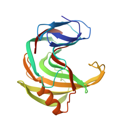
    6JUG, 6JWB, 6K9O, 6K9R, 6K9W, 6KW9, 6KWC, 6KWD, 6KWF, 6KWG

  • PubMed Abstract: 

    XynII is a family 11 glycoside hydrolase that uses the retaining mechanism for catalysis. In the active site, E177 works as the acid/base and E86 works as the nucleophile. Mutating an uncharged residue (N44) to an acidic residue (D) near E177 decreases the enzyme's optimal pH by ~ 1.0 unit. D44 was previously suggested to be a second proton carrier for catalysis. To test this hypothesis, we abolished the activity of E177 by mutating it to be Q, and mutated N44 to be D or E. These double mutants have dramatically decreased activities. Our high-resolution crystallographic structures and the microscopic pK a calculations show that D44 has similar position and pK a value during catalysis, indicating that D44 changes electrostatics around E177, which makes it prone to rotate as the acid/base in acidic conditions, thus decreases the pH optimum. Our results could be helpful to design enzymes with different pH optimum.


  • Organizational Affiliation
    • College of Science, Nanjing Agricultural University, Nanjing, 210095, People's Republic of China.

Macromolecule Content 

  • Total Structure Weight: 21.36 kDa 
  • Atom Count: 1,789 
  • Modeled Residue Count: 189 
  • Deposited Residue Count: 189 
  • Unique protein chains: 1

Macromolecules

Find similar proteins by:|  3D Structure
Entity ID: 1
MoleculeChains  Sequence LengthOrganismDetailsImage
Endo-1,4-beta-xylanase 2189Trichoderma reesei RUT C-30Mutation(s): 1 
Gene Names: xyn2
EC: 3.2.1.8
UniProt
Find proteins for P36217 (Hypocrea jecorina (strain ATCC 56765 / BCRC 32924 / NRRL 11460 / Rut C-30))
Explore P36217 
Go to UniProtKB:  P36217
Entity Groups
Sequence Clusters30% Identity50% Identity70% Identity90% Identity95% Identity100% Identity
UniProt GroupP36217
Sequence Annotations
Expand
Reference Sequence

Oligosaccharides

Help  
Entity ID: 2
MoleculeChains Length2D Diagram GlycosylationD Interactions
beta-D-xylopyranose-(1-4)-beta-D-xylopyranose-(1-4)-beta-D-xylopyranose
B
3N/A
Glycosylation Resources
GlyTouCan: G87429QT
GlyCosmos: G87429QT

Biologically Interesting Molecules (External Reference) 

1 Unique

Experimental Data & Validation

Experimental Data

  • Method: X-RAY DIFFRACTION
  • Resolution: 1.30 Å
  • R-Value Free:  0.163 (Depositor), 0.162 (DCC) 
  • R-Value Work:  0.150 (Depositor), 0.151 (DCC) 
  • R-Value Observed: 0.151 (Depositor) 
Space Group: P 21 21 21
Unit Cell:
Length ( Å )Angle ( ˚ )
a = 47.598α = 90
b = 59.161β = 90
c = 69.604γ = 90
Software Package:
Software NamePurpose
XDSdata reduction
SCALEPACKdata scaling
PHENIXrefinement
PDB_EXTRACTdata extraction
PHASERphasing

Structure Validation

View Full Validation Report


Ligand Structure Quality Assessment 


Entry History 

& Funding Information

Deposition Data

  • Released Date: 2020-12-30 
  • Deposition Author(s): Li, C., Wan, Q.

Funding OrganizationLocationGrant Number
National Natural Science Foundation of ChinaChina31670790

Revision History  (Full details and data files)

  • Version 1.0: 2020-12-30
    Type: Initial release
  • Version 1.1: 2023-11-22
    Changes: Data collection, Database references, Refinement description