


Crystal strcuture of PETase A248D, R280K mutant from Ideonella sakaiensis
Joo, S., Kim, K.-J.To be published.
Experimental Data Snapshot
Starting Model: experimental
View more details
wwPDB Validation 3D Report Full Report

Entity ID: 1 | |||||
|---|---|---|---|---|---|
| Molecule | Chains | Sequence Length | Organism | Details | Image |
| Poly(ethylene terephthalate) hydrolase | 300 | Pseudideonella sakaiensis | Mutation(s): 1 Gene Names: ISF6_4831 EC: 3.1.1.101 | 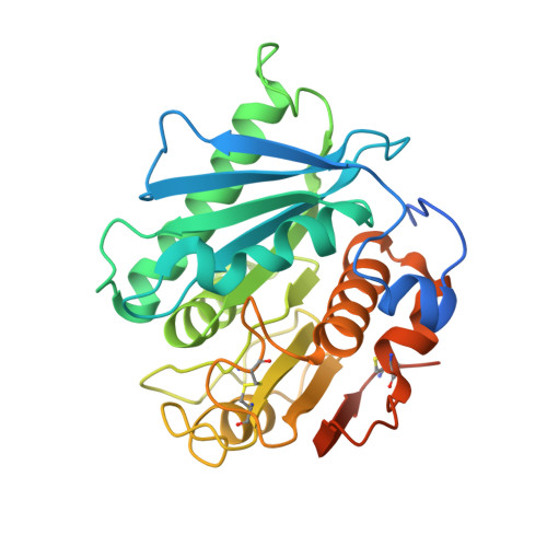 | |
UniProt | |||||
Find proteins for A0A0K8P6T7 (Piscinibacter sakaiensis) Explore A0A0K8P6T7 Go to UniProtKB: A0A0K8P6T7 | |||||
Entity Groups | |||||
| Sequence Clusters | 30% Identity50% Identity70% Identity90% Identity95% Identity100% Identity | ||||
| UniProt Group | A0A0K8P6T7 | ||||
Sequence AnnotationsExpand | |||||
Reference Sequence | |||||
| Length ( Å ) | Angle ( ˚ ) |
|---|---|
| a = 114.361 | α = 90 |
| b = 51.006 | β = 109.93 |
| c = 51.027 | γ = 90 |
| Software Name | Purpose |
|---|---|
| HKL-2000 | data reduction |
| REFMAC | refinement |
| PDB_EXTRACT | data extraction |
| HKL-2000 | data scaling |
| MOLREP | phasing |


