6KUB | pdb_00006kub

Crystal structure of Plasmodium falciparum histo-aspartic protease (HAP) zymogen (Form 1)


Experimental Data Snapshot

  • Method: X-RAY DIFFRACTION
  • Resolution: 2.00 Å
  • R-Value Free: 
    0.236 (Depositor), 0.247 (DCC) 
  • R-Value Work: 
    0.183 (Depositor) 
  • R-Value Observed: 
    0.186 (Depositor) 

Starting Model: experimental
View more details

wwPDB Validation 3D Report Full Report

Validation slider image for 6KUB

This is version 1.3 of the entry. See complete history

Literature

Activation mechanism of plasmepsins, pepsin-like aspartic proteases from Plasmodium, follows a unique trans-activation pathway.

Rathore, I.Mishra, V.Patel, C.Xiao, H.Gustchina, A.Wlodawer, A.Yada, R.Y.Bhaumik, P.

(2021) FEBS J 288: 678-698

  • DOI: https://doi.org/10.1111/febs.15363
  • Primary Citation Related Structures: 
    6KUB, 6KUC, 6KUD

  • PubMed Abstract: 

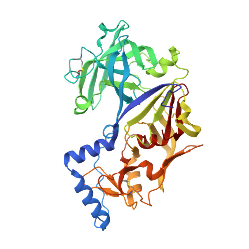
    Plasmodium parasites that cause malaria produce plasmepsins (PMs), pepsin-like aspartic proteases that are important antimalarial drug targets due to their role in host hemoglobin degradation. The enzymes are synthesized as inactive zymogens (pro-PMs), and the mechanism of their conversion to the active, mature forms has not been clearly elucidated. Our structural investigations of vacuolar pro-PMs with truncated prosegment (pro-tPMs) reveal that the formation of the S-shaped dimer is their innate property. Further structural studies, biochemical analysis, and molecular dynamics simulations indicate that disruption of the Tyr-Asp loop (121p-4), coordinated with the movement of the loop L1 (237-247) and helix H2 (101p-113p), is responsible for the extension of the pro-mature region (harboring the cleavage site). Consequently, under acidic pH conditions, these structural changes result in the dissociation of the dimers to monomers and the protonation of the residues in the prosegment prompts its unfolding. Subsequently, we demonstrated that the active site of the monomeric pro-tPMs with the unfolded prosegment is accessible for peptide substrate binding; in contrast, the active site is blocked in folded prosegment form of pro-tPMs. Thus, we propose a novel mechanism of auto-activation of vacuolar pro-tPMs that under acidic conditions can form a catalytically competent active site. One monomer cleaves the prosegment of the other one through a trans-activation process, resulting in formation of mature enzyme. As a result, once a mature enzyme is generated, it leads to the complete conversion of all the inactive pro-tPMs to their mature form. DATABASE: Atomic coordinates and structure factors have been submitted in the Protein Data Bank (PDB) under the PDB IDs 6KUB, 6KUC, and 6KUD.


  • Organizational Affiliation
    • Department of Biosciences and Bioengineering, Indian Institute of Technology Bombay, Mumbai, India.

Macromolecule Content 

  • Total Structure Weight: 43.92 kDa 
  • Atom Count: 3,477 
  • Modeled Residue Count: 380 
  • Deposited Residue Count: 380 
  • Unique protein chains: 1

Macromolecules

Find similar proteins by:|  3D Structure
Entity ID: 1
MoleculeChains  Sequence LengthOrganismDetailsImage
HAP protein380Plasmodium falciparumMutation(s): 0 
Gene Names: hap
EC: 3.4.23.39
UniProt
Find proteins for Q9Y006 (Plasmodium falciparum (isolate HB3))
Explore Q9Y006 
Go to UniProtKB:  Q9Y006
Entity Groups
Sequence Clusters30% Identity50% Identity70% Identity90% Identity95% Identity100% Identity
UniProt GroupQ9Y006
Sequence Annotations
Expand
Reference Sequence

Small Molecules

Ligands 2 Unique
IDChains Name / Formula / InChI Key2D Diagram3D Interactions
PEG

Query on PEG


Download:Ideal Coordinates CCD File
H [auth A],
I [auth A],
J [auth A]
DI(HYDROXYETHYL)ETHER
C4 H10 O3
MTHSVFCYNBDYFN-UHFFFAOYSA-N
GOL

Query on GOL


Download:Ideal Coordinates CCD File
B [auth A]
C [auth A]
D [auth A]
E [auth A]
F [auth A]
B [auth A],
C [auth A],
D [auth A],
E [auth A],
F [auth A],
G [auth A]
GLYCEROL
C3 H8 O3
PEDCQBHIVMGVHV-UHFFFAOYSA-N

Experimental Data & Validation

Experimental Data

  • Method: X-RAY DIFFRACTION
  • Resolution: 2.00 Å
  • R-Value Free:  0.236 (Depositor), 0.247 (DCC) 
  • R-Value Work:  0.183 (Depositor) 
  • R-Value Observed: 0.186 (Depositor) 
Space Group: C 1 2 1
Unit Cell:
Length ( Å )Angle ( ˚ )
a = 122.9α = 90
b = 69.3β = 126.1
c = 73.4γ = 90
Software Package:
Software NamePurpose
XSCALEdata scaling
REFMACrefinement
PDB_EXTRACTdata extraction
XDSdata reduction
PHASERphasing

Structure Validation

View Full Validation Report


Entry History 

& Funding Information

Deposition Data


Funding OrganizationLocationGrant Number
Department of Biotechnology (India)IndiaRamalingaswami re-entry fellowship

Revision History  (Full details and data files)

  • Version 1.0: 2020-05-27
    Type: Initial release
  • Version 1.1: 2021-02-03
    Changes: Database references
  • Version 1.2: 2023-11-22
    Changes: Data collection, Database references, Refinement description
  • Version 1.3: 2024-10-30
    Changes: Structure summary