On-Membrane Dynamic Interplay between Anti-GM1 IgG Antibodies and Complement Component C1q.
Yanaka, S., Yogo, R., Watanabe, H., Taniguchi, Y., Satoh, T., Komura, N., Ando, H., Yagi, H., Yuki, N., Uchihashi, T., Kato, K.(2019) Int J Mol Sci 21
- PubMed: 31878295 Search on PubMedSearch on PubMed Central
- DOI: https://doi.org/10.3390/ijms21010147
- Primary Citation Related Structures:
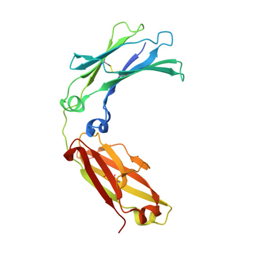
6KRU, 6KRV - PubMed Abstract:
Guillain-Barré syndrome, an autoimmune neuropathy characterized by acute limb weakness, is often preceded by Campylobacter jejuni infection. Molecular mimicry exists between the bacterial lipo-oligosaccharide and human ganglioside. Such C. jejuni infection induces production of immunoglobulin G1 (IgG1) autoantibodies against GM1 and causes complement-mediated motor nerve injury. For elucidating the molecular mechanisms linking autoantigen recognition and complement activation, we characterized the dynamic interactions of anti-GM1 IgG autoantibodies on ganglioside-incorporated membranes. Using high-speed atomic force microscopy, we found that the IgG molecules assemble into a hexameric ring structure on the membranes depending on their specific interactions with GM1. Complement component C1q was specifically recruited onto these IgG rings. The ring formation was inhibited by an IgG-binding domain of staphylococcal protein A bound at the cleft between the C H 2 and C H 3 domains. These data indicate that the IgG assembly is mediated through Fc-Fc interactions, which are promoted under on-membrane conditions due to restricted translational diffusion of IgG molecules. Reduction and alkylation of the hinge disulfide impaired IgG ring formation, presumably because of an increase in conformational entropic penalty. Our findings provide mechanistic insights into the molecular processes involved in Guillain-Barré syndrome and, more generally, into antigen-dependent interplay between antibodies and complement components on membranes.
- Exploratory Research Center on Life and Living Systems (ExCELLS) and Institute for Molecular Science (IMS), National Institutes of Natural Sciences, 5-1 Higashiyama, Myodaiji, Okazaki, Aichi 444-8787, Japan.
Organizational Affiliation: