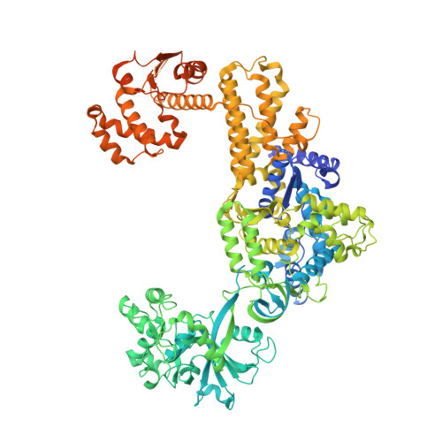
6KR7

Crystal structure of methylated human leucyl-tRNA synthetase, Leu-AMS-bound form


Experimental Data Snapshot

  • Method: X-RAY DIFFRACTION
  • Resolution: 4.00 Å
  • R-Value Free: 0.278 
  • R-Value Work: 0.232 
  • R-Value Observed: 0.237 

Starting Model: experimental
View more details

wwPDB Validation   3D Report Full Report


This is version 1.2 of the entry. See complete history


Literature

Leucine-sensing mechanism of leucyl-tRNA synthetase 1 for mTORC1 activation.

Kim, S.Yoon, I.Son, J.Park, J.Kim, K.Lee, J.H.Park, S.Y.Kang, B.S.Han, J.M.Hwang, K.Y.Kim, S.

(2021) Cell Rep 35: 109031-109031

  • DOI: https://doi.org/10.1016/j.celrep.2021.109031
  • Primary Citation of Related Structures:  
    6KID, 6KIE, 6KQY, 6KR7

  • PubMed Abstract: 

    Leucyl-tRNA synthetase 1 (LARS1) mediates activation of leucine-dependent mechanistic target of rapamycin complex 1 (mTORC1) as well as ligation of leucine to its cognate tRNAs, yet its mechanism of leucine sensing is poorly understood. Here we describe leucine binding-induced conformational changes of LARS1. We determine different crystal structures of LARS1 complexed with leucine, ATP, and a reaction intermediate analog, leucyl-sulfamoyl-adenylate (Leu-AMS), and find two distinct functional states of LARS1 for mTORC1 activation. Upon leucine binding to the synthetic site, H251 and R517 in the connective polypeptide and 50 FPYPY 54 in the catalytic domain change the hydrogen bond network, leading to conformational change in the C-terminal domain, correlating with RagD association. Leucine binding to LARS1 is increased in the presence of ATP, further augmenting leucine-dependent interaction of LARS1 and RagD. Thus, this work unveils the structural basis for leucine-dependent long-range communication between the catalytic and RagD-binding domains of LARS1 for mTORC1 activation.


  • Organizational Affiliation

    Department of Biotechnology, College of Life Sciences and Biotechnology, Korea University, Seoul 02841, Republic of Korea.


Macromolecules
Find similar proteins by:  (by identity cutoff)  |  3D Structure
Entity ID: 1
MoleculeChains Sequence LengthOrganismDetailsImage
Leucine--tRNA ligase, cytoplasmic1,188Homo sapiensMutation(s): 0 
Gene Names: LARSKIAA1352
EC: 6.1.1.4
UniProt & NIH Common Fund Data Resources
Find proteins for Q9P2J5 (Homo sapiens)
Explore Q9P2J5 
Go to UniProtKB:  Q9P2J5
PHAROS:  Q9P2J5
GTEx:  ENSG00000133706 
Entity Groups  
Sequence Clusters30% Identity50% Identity70% Identity90% Identity95% Identity100% Identity
UniProt GroupQ9P2J5
Sequence Annotations
Expand
  • Reference Sequence
Small Molecules
Binding Affinity Annotations 
IDSourceBinding Affinity
LSS BindingDB:  6KR7 IC50: 22 (nM) from 1 assay(s)
Experimental Data & Validation

Experimental Data

  • Method: X-RAY DIFFRACTION
  • Resolution: 4.00 Å
  • R-Value Free: 0.278 
  • R-Value Work: 0.232 
  • R-Value Observed: 0.237 
  • Space Group: P 65 2 2
Unit Cell:
Length ( Å )Angle ( ˚ )
a = 136.818α = 90
b = 136.818β = 90
c = 437.266γ = 120
Software Package:
Software NamePurpose
PHENIXrefinement
SCALEPACKdata scaling
PDB_EXTRACTdata extraction
HKL-2000data reduction
PHENIXphasing

Structure Validation

View Full Validation Report


Entry History 

Deposition Data

Revision History  (Full details and data files)

  • Version 1.0: 2021-01-27
    Type: Initial release
  • Version 1.1: 2021-08-11
    Changes: Database references
  • Version 1.2: 2023-11-22
    Changes: Data collection, Refinement description