


crystal structure of Xanthine-guanine phosphoribosyltransferase (XGPRT) from Yersinia pestis in P21212 space group
Lankipalli, S., Ramagopal, U.A.To be published.
Experimental Data Snapshot
Starting Model: experimental
View more details
Entity ID: 1 | |||||
|---|---|---|---|---|---|
| Molecule | Chains | Sequence Length | Organism | Details | Image |
| Xanthine phosphoribosyltransferase | 153 | Yersinia pestis | Mutation(s): 0 Gene Names: gpt, YPO3225, y0963, YP_0708 EC: 2.4.2.22 (PDB Primary Data), 2.4.2 (UniProt) | 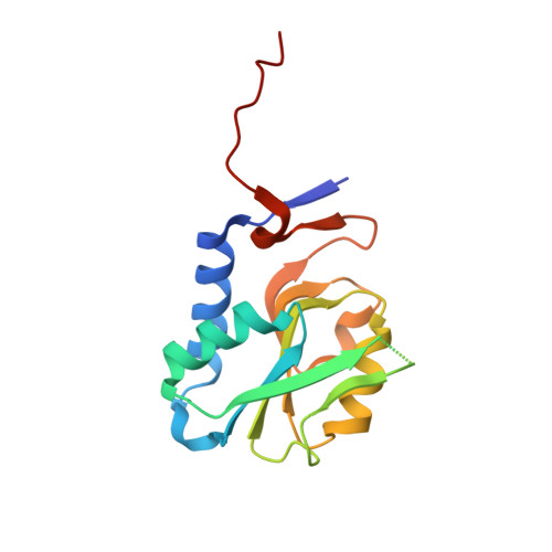 | |
UniProt | |||||
Entity Groups | |||||
| Sequence Clusters | 30% Identity50% Identity70% Identity90% Identity95% Identity100% Identity | ||||
| UniProt Group | Q8ZC05 | ||||
Sequence AnnotationsExpand | |||||
Reference Sequence | |||||
| Ligands 1 Unique | |||||
|---|---|---|---|---|---|
| ID | Chains | Name / Formula / InChI Key | 2D Diagram | 3D Interactions | |
| SO4 (Subject of Investigation/LOI) Download:Ideal Coordinates CCD File | C [auth A], D [auth A], E [auth B], F [auth B] | SULFATE ION O4 S QAOWNCQODCNURD-UHFFFAOYSA-L |  | ||
| Length ( Å ) | Angle ( ˚ ) |
|---|---|
| a = 58.32 | α = 90 |
| b = 94.63 | β = 90 |
| c = 51.67 | γ = 90 |
| Software Name | Purpose |
|---|---|
| REFMAC | refinement |
| Aimless | data scaling |
| PDB_EXTRACT | data extraction |
| iMOSFLM | data reduction |
| MOLREP | phasing |


