Primary Citation Related Structures: 6KKR, 6KKT, 6KKU
PubMed Abstract:
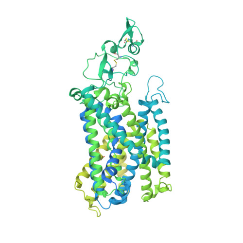
Cation-chloride cotransporters (CCCs) mediate the coupled, electroneutral symport of cations with chloride across the plasma membrane and are vital for cell volume regulation, salt reabsorption in the kidney, and γ-aminobutyric acid (GABA)-mediated modulation in neurons. Here we present cryo-electron microscopy (cryo-EM) structures of human potassium-chloride cotransporter KCC1 in potassium chloride or sodium chloride at 2.9- to 3.5-angstrom resolution. KCC1 exists as a dimer, with both extracellular and transmembrane domains involved in dimerization. The structural and functional analyses, along with computational studies, reveal one potassium site and two chloride sites in KCC1, which are all required for the ion transport activity. KCC1 adopts an inward-facing conformation, with the extracellular gate occluded. The KCC1 structures allow us to model a potential ion transport mechanism in KCCs and provide a blueprint for drug design.
Organizational Affiliation:
Department of Biophysics, Department of Pathology of Sir Run Run Shaw Hospital, Zhejiang University School of Medicine, Hangzhou 310058, China.
Tianjin Key Laboratory of Function and Application of Biological Macromolecular Structures, School of Life Sciences, Tianjin University, 92 Weijin Road, Nankai District, Tianjin 300072, China.
Center of Cryo-Electron Microscopy, Zhejiang University School of Medicine, Hangzhou 310058, China.
Zhejiang Province Key Laboratory of Quantum Technology and Device, Institute of Quantitative Biology, Department of Physics, Zhejiang University, Hangzhou, Zhejiang 310027, China.
Life Sciences Institute and Innovation Center for Cell Signaling Network, Zhejiang University, Hangzhou, Zhejiang 310058, China.
Department of Biophysics, Institute of Neuroscience, NHC and CAMS Key Laboratory of Medical Neurobiology, Zhejiang University School of Medicine, Hangzhou 310058, China.
Zhejiang Province Key Laboratory of Quantum Technology and Device, Institute of Quantitative Biology, Department of Physics, Zhejiang University, Hangzhou, Zhejiang 310027, China. jiangtaoguo@zju.edu.cn xiaochen.bai@utsouthwestern.edu sye@tju.edu.cn eric.delpire@vanderbilt.edu jingyuanli@zju.edu.cn.
Department of Anesthesiology, Vanderbilt University School of Medicine, Nashville, TN 37232, USA. jiangtaoguo@zju.edu.cn xiaochen.bai@utsouthwestern.edu sye@tju.edu.cn eric.delpire@vanderbilt.edu jingyuanli@zju.edu.cn.
Tianjin Key Laboratory of Function and Application of Biological Macromolecular Structures, School of Life Sciences, Tianjin University, 92 Weijin Road, Nankai District, Tianjin 300072, China. jiangtaoguo@zju.edu.cn xiaochen.bai@utsouthwestern.edu sye@tju.edu.cn eric.delpire@vanderbilt.edu jingyuanli@zju.edu.cn.
Departments of Biophysics and Cell Biology, University of Texas Southwestern Medical Center, Dallas, TX 75390, USA. jiangtaoguo@zju.edu.cn xiaochen.bai@utsouthwestern.edu sye@tju.edu.cn eric.delpire@vanderbilt.edu jingyuanli@zju.edu.cn.
Department of Biophysics, Department of Pathology of Sir Run Run Shaw Hospital, Zhejiang University School of Medicine, Hangzhou 310058, China. jiangtaoguo@zju.edu.cn xiaochen.bai@utsouthwestern.edu sye@tju.edu.cn eric.delpire@vanderbilt.edu jingyuanli@zju.edu.cn.