


Crystal structure of a two-quartet DNA G-quadruplex complexed with the porphyrin TMPyP4
Zhang, Y.S., Parkinson, G.N., Wei, D.G.To be published.
Experimental Data Snapshot
Starting Model: experimental
View more details
Entity ID: 1 | ||||
| Molecule | Chains | Length | Organism | Image |
|---|---|---|---|---|
| DNA (5'-D(*GP*GP*CP*TP*CP*GP*GP*CP*GP*GP*CP*GP*GP*A)-3') | 14 | Pseudorabies virus Ea | 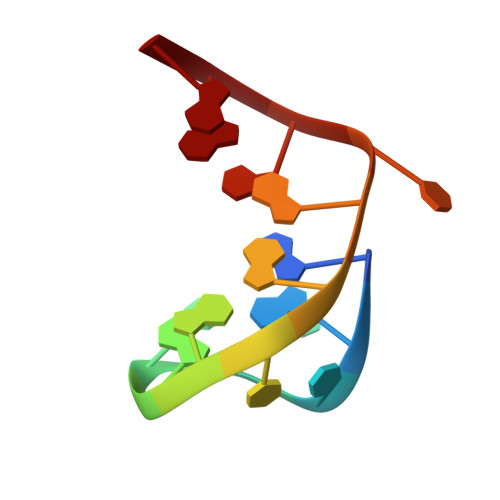 | |
Sequence AnnotationsExpand | ||||
Reference Sequence | ||||
| Ligands 3 Unique | |||||
|---|---|---|---|---|---|
| ID | Chains | Name / Formula / InChI Key | 2D Diagram | 3D Interactions | |
| POH (Subject of Investigation/LOI) Download:Ideal Coordinates CCD File | G [auth A] | (1Z,4Z,9Z,15Z)-5,10,15,20-tetrakis(1-methylpyridin-1-ium-4-yl)-21,23-dihydroporphyrin C44 H38 N8 ABCGFHPGHXSVKI-LWQDQPMZSA-O |  | ||
| NCO (Subject of Investigation/LOI) Download:Ideal Coordinates CCD File | H [auth A] | COBALT HEXAMMINE(III) Co H18 N6 DYLMFCCYOUSRTK-UHFFFAOYSA-N |  | ||
| K (Subject of Investigation/LOI) Download:Ideal Coordinates CCD File | C [auth A], D [auth A], E [auth A], F [auth A] | POTASSIUM ION K NPYPAHLBTDXSSS-UHFFFAOYSA-N |  | ||
| Length ( Å ) | Angle ( ˚ ) |
|---|---|
| a = 48.152 | α = 90 |
| b = 47.192 | β = 113.15 |
| c = 38.133 | γ = 90 |
| Software Name | Purpose |
|---|---|
| PHENIX | refinement |
| PDB_EXTRACT | data extraction |
| HKL-3000 | data reduction |
| HKL-3000 | data scaling |
| PHASER | phasing |
| Funding Organization | Location | Grant Number |
|---|---|---|
| National Natural Science Foundation of China | China | 31672558 |


