


Structural and catalytic analysis of two diverse uridine phosphorylases in the oomycete Phytophthora capsici.
Yang, C.C., Zhang, X.G.To be published.
Experimental Data Snapshot
wwPDB Validation 3D Report Full Report

Entity ID: 1 | |||||
|---|---|---|---|---|---|
| Molecule | Chains | Sequence Length | Organism | Details | Image |
| Uridine phosphorylase | 322 | Phytophthora capsici LT1534 | Mutation(s): 0 EC: 2.4.2.3 | 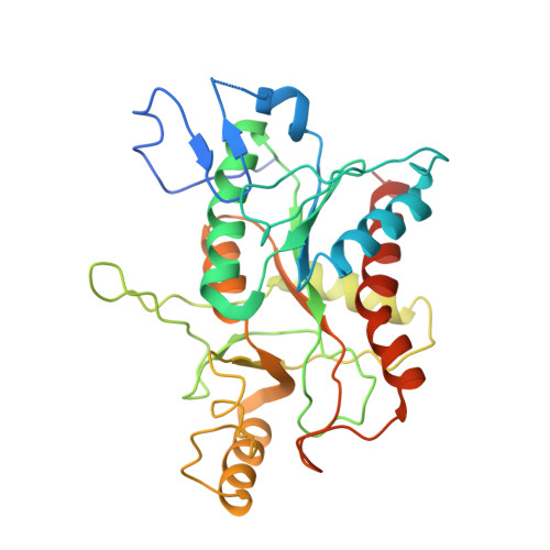 | |
| Length ( Å ) | Angle ( ˚ ) |
|---|---|
| a = 105.836 | α = 90 |
| b = 66.386 | β = 96.77 |
| c = 261.061 | γ = 90 |
| Software Name | Purpose |
|---|---|
| PHENIX | refinement |
| PDB_EXTRACT | data extraction |
| HKL-3000 | data reduction |
| HKL-3000 | data scaling |
| SOLVE | phasing |


