6K9T

Crystal structure of a class C beta-lactamase in complex with cefotaxime


Experimental Data Snapshot

  • Method: X-RAY DIFFRACTION
  • Resolution: 1.46 Å
  • R-Value Free: 0.190 
  • R-Value Work: 0.169 
  • R-Value Observed: 0.170 

wwPDB Validation   3D Report Full Report


Ligand Structure Quality Assessment 


This is version 1.2 of the entry. See complete history


Literature

Structural Insights into Catalytic Relevances of Substrate Poses in ACC-1.

Bae, D.W.Jung, Y.E.An, Y.J.Na, J.H.Cha, S.S.

(2019) Antimicrob Agents Chemother 63

  • DOI: https://doi.org/10.1128/AAC.01411-19
  • Primary Citation of Related Structures:  
    6K8X, 6K9T, 6KA5, 6KBY

  • PubMed Abstract: 

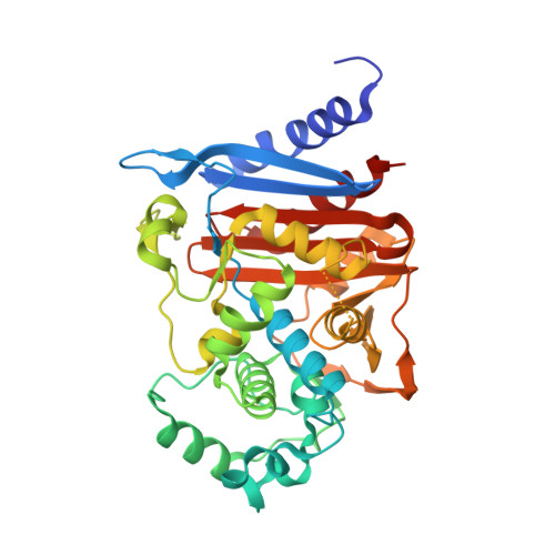
    ACC-1 is a plasmid-encoded class C β-lactamase identified in clinical isolates of Klebsiella pneumoniae , Proteus mirabilis , Salmonella enterica , and Escherichia coli ACC-1-producing bacteria are susceptible to cefoxitin, whereas they are resistant to oxyimino cephalosporins. Here, we depict crystal structures of apo ACC-1, adenylylated ACC-1, and acylated ACC-1 complexed with cefotaxime and cefoxitin. ACC-1 has noteworthy structural alterations in the R2 loop, the Ω loop, and the Phe119 loop located along the active-site rim. The adenylate covalently bonded to the nucleophilic serine reveals a tetrahedral phosphorus mimicking the deacylation transition state. Cefotaxime in ACC-1 has a proper conformation for the substrate-assisted catalysis in that its C-4 carboxylate and N-5 nitrogen are adequately located to facilitate the deacylation reaction. In contrast, cefoxitin in ACC-1 has a distinct conformation, in which those functional groups cannot contribute to catalysis. Furthermore, the orientation of the deacylating water relative to the acyl carbonyl group in ACC-1 is unfavorable for nucleophilic attack.


  • Organizational Affiliation

    Department of Chemistry & Nanoscience, Ewha Womans University, Seoul, Republic of Korea.


Macromolecules
Find similar proteins by:  (by identity cutoff)  |  3D Structure
Entity ID: 1
MoleculeChains Sequence LengthOrganismDetailsImage
Beta-lactamase370Klebsiella pneumoniaeMutation(s): 0 
Gene Names: bla-ACC-1acc-1
EC: 3.5.2.6
UniProt
Find proteins for Q9XB24 (Klebsiella pneumoniae)
Explore Q9XB24 
Go to UniProtKB:  Q9XB24
Entity Groups  
Sequence Clusters30% Identity50% Identity70% Identity90% Identity95% Identity100% Identity
UniProt GroupQ9XB24
Sequence Annotations
Expand
  • Reference Sequence
Small Molecules
Ligands 1 Unique
IDChains Name / Formula / InChI Key2D Diagram3D Interactions
CEF (Subject of Investigation/LOI)
Query on CEF

Download Ideal Coordinates CCD File 
B [auth A]CEFOTAXIME, C3' cleaved, open, bound form
C14 H15 N5 O5 S2
NRYMPLKBKFIWQC-YVCCLBOHSA-N
Experimental Data & Validation

Experimental Data

  • Method: X-RAY DIFFRACTION
  • Resolution: 1.46 Å
  • R-Value Free: 0.190 
  • R-Value Work: 0.169 
  • R-Value Observed: 0.170 
  • Space Group: C 1 2 1
Unit Cell:
Length ( Å )Angle ( ˚ )
a = 133.219α = 90
b = 60.416β = 111.98
c = 56.039γ = 90
Software Package:
Software NamePurpose
PHENIXrefinement
HKL-2000data scaling
PDB_EXTRACTdata extraction
Cootmodel building
HKL-2000data reduction

Structure Validation

View Full Validation Report


Ligand Structure Quality Assessment 


Entry History 

Deposition Data

Revision History  (Full details and data files)

  • Version 1.0: 2019-10-16
    Type: Initial release
  • Version 1.1: 2019-11-06
    Changes: Data collection, Database references
  • Version 1.2: 2024-03-20
    Changes: Data collection, Database references, Refinement description