Crystal contact-free conformation of an intrinsically flexible loop in protein crystal: Tim21 as the case study.
Bala, S., Shinya, S., Srivastava, A., Ishikawa, M., Shimada, A., Kobayashi, N., Kojima, C., Tama, F., Miyashita, O., Kohda, D.(2020) Biochim Biophys Acta Gen Subj 1864: 129418-129418
- PubMed: 31449839 Search on PubMed
- DOI: https://doi.org/10.1016/j.bbagen.2019.129418
- Primary Citation Related Structures:
6K7D, 6K7E, 6K7F, 6K8Q - PubMed Abstract:
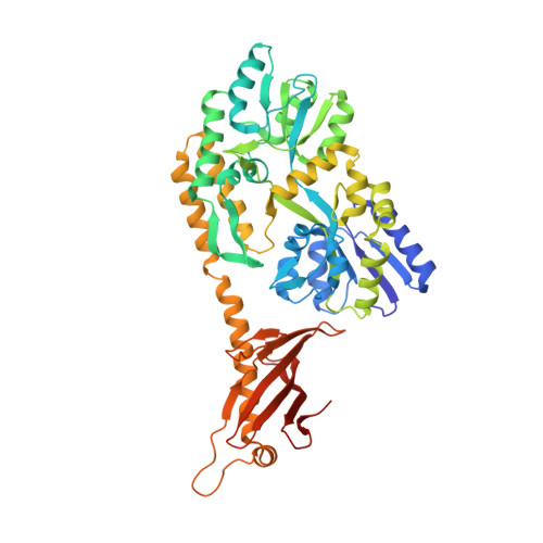
In protein crystals, flexible loops are frequently deformed by crystal contacts, whereas in solution, the large motions result in the poor convergence of such flexible loops in NMR structure determinations. We need an experimental technique to characterize the structural and dynamic properties of intrinsically flexible loops of protein molecules. We designed an intended crystal contact-free space (CCFS) in protein crystals, and arranged the flexible loop of interest in the CCFS. The yeast Tim 21 protein was chosen as the model protein, because one of the loops (loop 2) is distorted by crystal contacts in the conventional crystal. Yeast Tim21 was fused to the MBP protein by a rigid α-helical linker. The space created between the two proteins was used as the CCFS. The linker length provides adjustable freedom to arrange loop 2 in the CCFS. We re-determined the NMR structure of yeast Tim21, and conducted MD simulations for comparison. Multidimensional scaling was used to visualize the conformational similarity of loop 2. We found that the crystal contact-free conformation of loop 2 is located close to the center of the ensembles of the loop 2 conformations in the NMR and MD structures. Loop 2 of yeast Tim21 in the CCFS adopts a representative, dominant conformation in solution. No single powerful technique is available for the characterization of flexible structures in protein molecules. NMR analyses and MD simulations provide useful, but incomplete information. CCFS crystallography offers a third route to this goal.
- Division of Structural Biology, Medical Institute of Bioregulation, Kyushu University, Maidashi 3-1-1, Higashi-ku, Fukuoka 812-8582, Japan.
Organizational Affiliation: