Unique active site formation in a novel galactose 1-phosphate uridylyltransferase from the hyperthermophilic archaeon Pyrobaculum aerophilum.
Ohshida, T., Hayashi, J., Yoneda, K., Ohshima, T., Sakuraba, H.(2020) Proteins 88: 669-678
- PubMed: 31693208 Search on PubMed
- DOI: https://doi.org/10.1002/prot.25848
- Primary Citation Related Structures:
6K5Z, 6K9Z - PubMed Abstract:
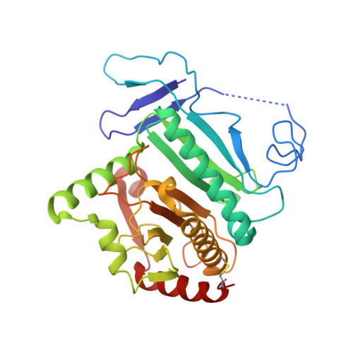
A gene encoding galactose 1-phosphate uridylyltransferase (GalT) was identified in the hyperthermophilic archaeon Pyrobaculum aerophilum. The gene was overexpressed in Escherichia coli, after which its product was purified and characterized. The expressed enzyme was highly thermostable and retained about 90% of its activity after incubation for 10 minutes at temperatures up to 90°C. Two different crystal structures of P. aerophilum GalT were determined: the substrate-free enzyme at 2.33 Å and the UDP-bound H140F mutant enzyme at 1.78 Å. The main-chain coordinates of the P. aerophilum GalT monomer were similar to those in the structures of the E. coli and human GalTs, as was the dimeric arrangement. However, there was a striking topological difference between P. aerophilum GalT and the other two enzymes. In the E. coli and human enzymes, the N-terminal chain extends from one subunit into the other and forms part of the substrate-binding pocket in the neighboring subunit. By contrast, the N-terminal chain in P. aerophilum GalT extends to the substrate-binding site in the same subunit. Amino acid sequence alignment showed that a shorter surface loop in the N-terminal region contributes to the unique topology of P. aerophilum GalT. Structural comparison of the substrate-free enzyme with UDP-bound H140F suggests that binding of the glucose moiety of the substrate, but not the UDP moiety, gives rise to a large structural change around the active site. This may in turn provide an appropriate environment for the enzyme reaction.
- Department of Applied Biological Science, Faculty of Agriculture, Kagawa University, Miki-cho, Kagawa, Japan.
Organizational Affiliation: