6K5F | pdb_00006k5f

Crystal structure of the CLC-ec1 deltaNC in presence of 200 mM NaBr


Experimental Data Snapshot

  • Method: X-RAY DIFFRACTION
  • Resolution: 3.20 Å
  • R-Value Free: 
    0.300 (Depositor), 0.316 (DCC) 
  • R-Value Work: 
    0.236 (Depositor), 0.254 (DCC) 
  • R-Value Observed: 
    0.239 (Depositor) 

Starting Model: experimental
View more details

wwPDB Validation 3D Report Full Report

Validation slider image for 6K5F

This is version 1.4 of the entry. See complete history

Literature

Mutation of external glutamate residue reveals a new intermediate transport state and anion binding site in a CLC Cl-/H+antiporter.

Park, K.Lee, B.C.Lim, H.H.

(2019) Proc Natl Acad Sci U S A 116: 17345-17354

  • DOI: https://doi.org/10.1073/pnas.1901822116
  • Primary Citation Related Structures: 
    6AD7, 6AD8, 6ADA, 6ADB, 6ADC, 6K5A, 6K5D, 6K5F, 6K5I

  • PubMed Abstract: 

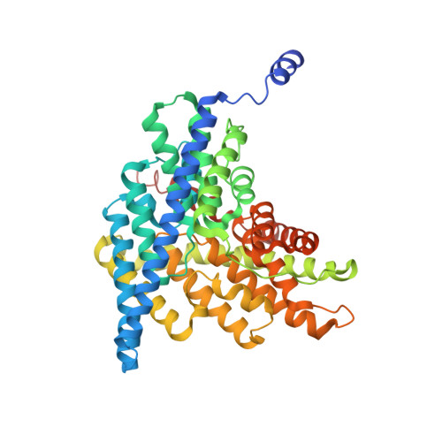
    The CLC family of proteins are involved in a variety of physiological processes to control cellular chloride concentration. Two distinct classes of CLC proteins, Cl - channels and Cl - /H + antiporters, have been functionally and structurally investigated over the last several decades. Previous studies have suggested that the conformational heterogeneity of the critical glutamate residue, Glu ex , could explain the transport cycle of CLC-type Cl - /H + antiporters. However, the presence of multiple conformations ( Up , Middle , and Down ) of the Glu ex has been suggested from combined structural snapshots of 2 different CLC antiporters: CLC-ec1 from Escherichia coli and cmCLC from a thermophilic red alga, Cyanidioschyzon merolae Thus, we aimed to investigate further the heterogeneity of Glu ex -conformations in CLC-ec1, the most deeply studied CLC antiporter, at both functional and structural levels. Here, we show that the crystal structures of the Glu ex mutant E148D and wild-type CLC-ec1 with varying anion concentrations suggest a structural intermediate, the " Midlow " conformation. We also found that an extra anion can be located above the external Cl - -binding site in the E148D mutant when the anion concentration is high. Moreover, we observed that a carboxylate in solution can occupy either the external or central Cl - -binding site in the ungated E148A mutant using an anomalously detectable short carboxylic acid, bromoacetate. These results lend credibility to the idea that the Glu ex can take at least 3 distinct conformational states during the transport cycle of a single CLC antiporter.


  • Organizational Affiliation
    • Department of Structure and Function of Neural Network, Korea Brain Research Institute, 41068 Daegu, Republic of Korea.

Macromolecule Content 

  • Total Structure Weight: 194.92 kDa 
  • Atom Count: 13,247 
  • Modeled Residue Count: 1,751 
  • Deposited Residue Count: 1,812 
  • Unique protein chains: 3

Macromolecules

Find similar proteins by:|  3D Structure
Entity ID: 1
MoleculeChains  Sequence LengthOrganismDetailsImage
H(+)/Cl(-) exchange transporter ClcA
A, B
473Escherichia coliMutation(s): 0 
Gene Names: 
yadQclcAeriCA6592_03060A9819_00720A9R57_04335AC067_04570AC789_1c01550ACN002_0160ACN81_28610ACU57_18640AM270_18100AM446_22830AMK83_01920AWP75_13405B1K96_13065B7C53_01430B9M99_16180BANRA_02286BANRA_02303BANRA_03326BB545_20825BE963_14760BER14_07860BFD29_18565BHF46_07945BHS81_00575BHS87_00725BIQ87_00715BIU72_13480BJJ90_21425BK292_13605BK373_09530BK383_20300BMT53_09575BMT91_01430BN17_45791BVL39_02260BXT93_20950BZL31_15015BZL69_20275C2M16_10720C2U48_09685C3444_11105C3449_15895C4J69_03510C5715_17420C5P01_08850C5P43_19885C7235_20305C7B02_09235CG691_14570CIJ94_20900COD46_06560CR538_20715CRD98_15250CRM83_15165CRT46_00820CV83915_01419CWM24_04960CWS33_09545CXB56_23210D2184_08665D2185_20055D2188_12715D3821_06135D3822_17405D3I61_01530D3O91_03110D3Y67_10980D5618_10265D5653_16505D8K42_10345D9D20_18555D9D29_16860D9D31_08820D9D55_03155D9D69_06945D9E22_13175D9E34_01170D9F17_00235D9F57_04830D9H66_18270D9H68_13700D9H94_13970D9I47_04165D9I87_01705D9I88_06500D9I97_03880D9J03_00240D9J11_16750D9J44_14145D9J60_02140DL545_20465DLW18_08025DMZ31_06935DNQ26_06330DP277_13800DQF57_05740DQO13_06325DS732_05700DTL43_06000DTM10_02705DTM45_12560DU321_03585EAI42_18265EAI44_17040EAI46_14565EAI52_10940EC1094V2_3697EC3234A_2c01370Eco118UI_00790EEP23_00240EFV01_09785EFV02_20350EFV04_15100EFV08_17265EFV11_08020EFV12_18730EFV14_08750EFV15_17340EFV17_09990EIA10_05995EL75_3609EL79_3718EL80_3665EOL26_04835ERS085366_02706ERS085379_00551ERS150873_01097GJ11_00825HMPREF3040_02596MJ49_06260NCTC10082_01396NCTC10090_02173NCTC10429_04214NCTC11126_03619NCTC12950_04437NCTC13148_05172NCTC7922_05796NCTC7927_04520NCTC8009_07339NCTC8179_01364NCTC8500_04562NCTC8959_04830NCTC9036_04037NCTC9037_04203NCTC9044_03171NCTC9050_02039NCTC9055_00980NCTC9058_02783NCTC9062_04200NCTC9073_03413NCTC9077_05114NCTC9111_04278NCTC9702_04777NCTC9703_03448NCTC9706_01351NCTC9777_00485PU06_23100RG28_03970RK56_028075RX35_04890SAMEA3472044_04171SAMEA3472055_02614SAMEA3472070_02408SAMEA3472090_04472SAMEA3472110_04526SAMEA3472112_04666SAMEA3472147_00367SAMEA3484427_01361SAMEA3484429_01120SAMEA3484433_03082SAMEA3485113_00827SAMEA3752372_04445SAMEA3752553_02908SAMEA3752557_00860SAMEA3752559_03726SAMEA3752620_02075SAMEA3753097_03891SAMEA3753164_02448SK85_00155UC41_07360UN86_07645WQ89_09495WR15_02265YDC107_3382

Membrane Entity: Yes 
UniProt
Find proteins for P37019 (Escherichia coli (strain K12))
Explore P37019 
Go to UniProtKB:  P37019
Entity Groups
Sequence Clusters30% Identity50% Identity70% Identity90% Identity95% Identity100% Identity
UniProt GroupP37019
Sequence Annotations
Expand
Reference Sequence
Find similar proteins by:|  3D Structure
Entity ID: 2
MoleculeChains  Sequence LengthOrganismDetailsImage
Fab fragment, heavy chain
C, E
222Mus musculusMutation(s): 0 
Find similar proteins by:|  3D Structure
Entity ID: 3
MoleculeChains  Sequence LengthOrganismDetailsImage
Fab fragment, light chain
D, F
211Mus musculusMutation(s): 0 

Experimental Data & Validation

Experimental Data

  • Method: X-RAY DIFFRACTION
  • Resolution: 3.20 Å
  • R-Value Free:  0.300 (Depositor), 0.316 (DCC) 
  • R-Value Work:  0.236 (Depositor), 0.254 (DCC) 
  • R-Value Observed: 0.239 (Depositor) 
Space Group: C 1 2 1
Unit Cell:
Length ( Å )Angle ( ˚ )
a = 230.789α = 90
b = 101.656β = 132.09
c = 172.003γ = 90
Software Package:
Software NamePurpose
PHENIXrefinement
HKL-2000data reduction
HKL-2000data scaling
PHASERphasing

Structure Validation

View Full Validation Report


Entry History 

& Funding Information

Deposition Data


Funding OrganizationLocationGrant Number
Ministry of Science, ICT and Future PlanningKorea, Republic Of19-BR-01-02
Ministry of Science, ICT and Future PlanningKorea, Republic Of2017M3C7A 1048086

Revision History  (Full details and data files)

  • Version 1.0: 2019-08-28
    Type: Initial release
  • Version 1.1: 2019-09-11
    Changes: Data collection, Database references
  • Version 1.2: 2019-11-13
    Changes: Source and taxonomy
  • Version 1.3: 2023-11-22
    Changes: Data collection, Database references, Refinement description
  • Version 1.4: 2024-11-06
    Changes: Structure summary