High-resolution structures of annexin A5 in a two-dimensional array.
Hong, S., Na, S., Kim, O.H., Jeong, S., Oh, B.C., Ha, N.C.(2020) J Struct Biol 209: 107401-107401
- PubMed: 31605770 Search on PubMed
- DOI: https://doi.org/10.1016/j.jsb.2019.10.003
- Primary Citation Related Structures:
6K22, 6K25 - PubMed Abstract:
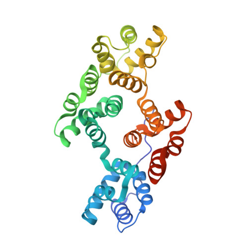
Annexins are soluble cytosolic proteins that bind to cell membranes. Annexin A5 self-assembles into a two-dimensional (2D) array and prevents cell rupture by attaching to damaged membranes. However, this process is not fully understood at the molecular level. In this study, we determined the crystal structures of annexin A5 with and without calcium (Ca 2+ ) and confirmed the Ca 2+ -dependent outward motion of a tryptophan residue. Strikingly, the two structures exhibited the same crystal packing and 2D arrangement into a p3 lattice, which agrees well with the results of low-resolution structural imaging. High-resolution structures indicated that a three-fold interaction near the tryptophan residue is important for mediating the formation of the p3 lattice. A hypothesis on the promotion of p3 lattice formation by phosphatidyl serine (PS) is also suggested. This study provides molecular insight into how annexins modulate the physical properties of cell membranes as a function of Ca 2+ concentration and the phospholipid composition of the membrane.
- Department of Agricultural Biotechnology, Research Institute of Agriculture and Life Sciences, Center for Food and Bioconvergence, Center for Food Safety and Toxicology, Seoul National University, Seoul 08826, Republic of Korea.
Organizational Affiliation: