6JWA | pdb_00006jwa

Crystal structure of CK2a1 with 5-iodotubercidin


Experimental Data Snapshot

  • Method: X-RAY DIFFRACTION
  • Resolution: 1.78 Å
  • R-Value Free: 
    0.225 (Depositor), 0.225 (DCC) 
  • R-Value Work: 
    0.185 (Depositor), 0.187 (DCC) 
  • R-Value Observed: 
    0.187 (Depositor) 

Starting Model: experimental
View more details

wwPDB Validation 3D Report Full Report

Validation slider image for 6JWA

Ligand Structure Quality Assessment 


This is version 1.1 of the entry. See complete history

Literature

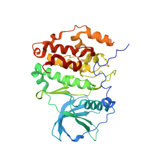
A promiscuous kinase inhibitor delineates the conspicuous structural features of protein kinase CK2a1.

Tsuyuguchi, M.Nakaniwa, T.Sawa, M.Nakanishi, I.Kinoshita, T.

(2019) Acta Crystallogr F Struct Biol Commun 75: 515-519

  • DOI: https://doi.org/10.1107/S2053230X19008951
  • Primary Citation Related Structures: 
    6JWA

  • PubMed Abstract: 

    Protein kinase CK2a1 is a serine/threonine kinase that plays a crucial role in the growth, proliferation and survival of cells and is a well known target for tumour and glomerulonephritis therapies. Here, the crystal structure of the kinase domain of CK2a1 complexed with 5-iodotubercidin (5IOD), an ATP-mimetic inhibitor, was determined at 1.78 Å resolution. The structure shows distinct structural features and, in combination with a comparison of the crystal structures of five off-target kinases complexed with 5IOD, provides valuable information for the development of highly selective inhibitors.


  • Organizational Affiliation
    • Graduate School of Science, Osaka Prefecture University, 1-1 Gakuen-cho, Naka-ku, Sakai, Osaka 599-8531, Japan.

Macromolecule Content 

  • Total Structure Weight: 41.24 kDa 
  • Atom Count: 2,963 
  • Modeled Residue Count: 327 
  • Deposited Residue Count: 340 
  • Unique protein chains: 1

Macromolecules

Find similar proteins by:|  3D Structure
Entity ID: 1
MoleculeChains  Sequence LengthOrganismDetailsImage
Casein kinase II subunit alpha340Homo sapiensMutation(s): 0 
Gene Names: CSNK2A1CK2A1
EC: 2.7.11.1
UniProt & NIH Common Fund Data Resources
Find proteins for P68400 (Homo sapiens)
Explore P68400 
Go to UniProtKB:  P68400
PHAROS:  P68400
GTEx:  ENSG00000101266 
Entity Groups
Sequence Clusters30% Identity50% Identity70% Identity90% Identity95% Identity100% Identity
UniProt GroupP68400
Sequence Annotations
Expand
Reference Sequence

Small Molecules

Ligands 2 Unique
IDChains Name / Formula / InChI Key2D Diagram3D Interactions
5ID
(Subject of Investigation/LOI)

Query on 5ID


Download:Ideal Coordinates CCD File
B [auth A](2R,3R,4S,5R)-2-(4-AMINO-5-IODO-7H-PYRROLO[2,3-D]PYRIMIDIN-7-YL)-5-(HYDROXYMETHYL)TETRAHYDROFURAN-3,4-DIOL
C11 H13 I N4 O4
WHSIXKUPQCKWBY-IOSLPCCCSA-N
EDO

Query on EDO


Download:Ideal Coordinates CCD File
C [auth A]
D [auth A]
E [auth A]
F [auth A]
G [auth A]
C [auth A],
D [auth A],
E [auth A],
F [auth A],
G [auth A],
H [auth A]
1,2-ETHANEDIOL
C2 H6 O2
LYCAIKOWRPUZTN-UHFFFAOYSA-N

Experimental Data & Validation

Experimental Data

  • Method: X-RAY DIFFRACTION
  • Resolution: 1.78 Å
  • R-Value Free:  0.225 (Depositor), 0.225 (DCC) 
  • R-Value Work:  0.185 (Depositor), 0.187 (DCC) 
  • R-Value Observed: 0.187 (Depositor) 
Space Group: P 21 21 21
Unit Cell:
Length ( Å )Angle ( ˚ )
a = 48.21α = 90
b = 78.936β = 90
c = 82.842γ = 90
Software Package:
Software NamePurpose
PHENIXrefinement
HKL-2000data reduction
HKL-2000data scaling
PHASERphasing

Structure Validation

View Full Validation Report


Ligand Structure Quality Assessment 


Entry History 

Deposition Data

Revision History  (Full details and data files)

  • Version 1.0: 2019-07-24
    Type: Initial release
  • Version 1.1: 2023-11-22
    Changes: Data collection, Database references, Refinement description