6JP5 | pdb_00006jp5

Rabbit Cav1.1-Nifedipine Complex


Experimental Data Snapshot

  • Method: ELECTRON MICROSCOPY
  • Resolution: 2.90 Å
  • Aggregation State: PARTICLE 
  • Reconstruction Method: SINGLE PARTICLE 

wwPDB Validation 3D Report Full Report

Validation slider image for 6JP5

This is version 3.1 of the entry. See complete history

Literature

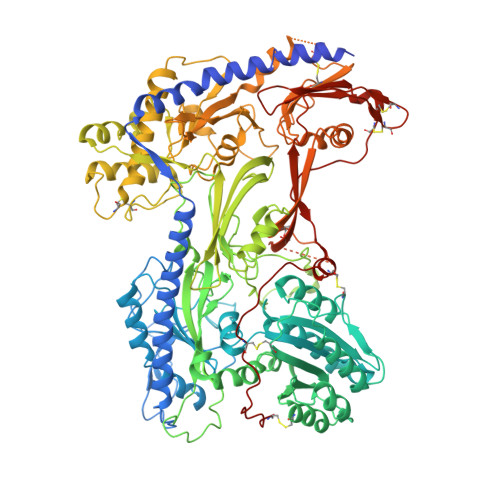
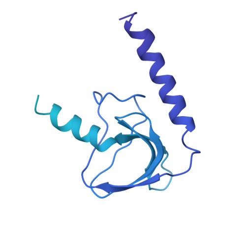
Molecular Basis for Ligand Modulation of a Mammalian Voltage-Gated Ca2+Channel.

Zhao, Y.Huang, G.Wu, J.Wu, Q.Gao, S.Yan, Z.Lei, J.Yan, N.

(2019) Cell 177: 1495-1506.e12

  • DOI: https://doi.org/10.1016/j.cell.2019.04.043
  • Primary Citation Related Structures: 
    6JP5, 6JP8, 6JPA, 6JPB

  • PubMed Abstract: 

    The L-type voltage-gated Ca 2+ (Ca v ) channels are modulated by various compounds exemplified by 1,4-dihydropyridines (DHP), benzothiazepines (BTZ), and phenylalkylamines (PAA), many of which have been used for characterizing channel properties and for treatment of hypertension and other disorders. Here, we report the cryoelectron microscopy (cryo-EM) structures of Ca v 1.1 in complex with archetypal antagonistic drugs, nifedipine, diltiazem, and verapamil, at resolutions of 2.9 Å, 3.0 Å, and 2.7 Å, respectively, and with a DHP agonist Bay K 8644 at 2.8 Å. Diltiazem and verapamil traverse the central cavity of the pore domain, directly blocking ion permeation. Although nifedipine and Bay K 8644 occupy the same fenestration site at the interface of repeats III and IV, the coordination details support previous functional observations that Bay K 8644 is less favored in the inactivated state. These structures elucidate the modes of action of different Ca v ligands and establish a framework for structure-guided drug discovery.


  • Organizational Affiliation
    • State Key Laboratory of Membrane Biology, Beijing Advanced Innovation Center for Structural Biology, Tsinghua-Peking Joint Center for Life Sciences, School of Life Sciences, Tsinghua University, Beijing 100084, China.

Macromolecule Content 

  • Total Structure Weight: 470.87 kDa 
  • Atom Count: 22,068 
  • Modeled Residue Count: 2,694 
  • Deposited Residue Count: 4,068 
  • Unique protein chains: 4

Macromolecules

Find similar proteins by:|  3D Structure
Entity ID: 1
MoleculeChains  Sequence LengthOrganismDetailsImage
Voltage-dependent L-type calcium channel subunit alpha-1S1,873Oryctolagus cuniculusMutation(s): 0 
Membrane Entity: Yes 
UniProt
Find proteins for P07293 (Oryctolagus cuniculus)
Explore P07293 
Go to UniProtKB:  P07293
Entity Groups
Sequence Clusters30% Identity50% Identity70% Identity90% Identity95% Identity100% Identity
UniProt GroupP07293
Glycosylation
Glycosylation Sites: 1
Sequence Annotations
Expand
Reference Sequence
Find similar proteins by:|  3D Structure
Entity ID: 2
MoleculeChains  Sequence LengthOrganismDetailsImage
Voltage-dependent calcium channel gamma-1 subunitB [auth E]222Oryctolagus cuniculusMutation(s): 0 
Membrane Entity: Yes 
UniProt
Find proteins for P19518 (Oryctolagus cuniculus)
Explore P19518 
Go to UniProtKB:  P19518
Entity Groups
Sequence Clusters30% Identity50% Identity70% Identity90% Identity95% Identity100% Identity
UniProt GroupP19518
Sequence Annotations
Expand
Reference Sequence
Find similar proteins by:|  3D Structure
Entity ID: 3
MoleculeChains  Sequence LengthOrganismDetailsImage
Voltage-dependent calcium channel subunit alpha-2/delta-1C [auth F]1,073Oryctolagus cuniculusMutation(s): 0 
Membrane Entity: Yes 
UniProt
Find proteins for P13806 (Oryctolagus cuniculus)
Explore P13806 
Go to UniProtKB:  P13806
Entity Groups
Sequence Clusters30% Identity50% Identity70% Identity90% Identity95% Identity100% Identity
UniProt GroupP13806
Glycosylation
Glycosylation Sites: 8
Sequence Annotations
Expand
Reference Sequence
Find similar proteins by:|  3D Structure
Entity ID: 4
MoleculeChains  Sequence LengthOrganismDetailsImage
Voltage-dependent L-type calcium channel subunit beta-1D [auth B],
E [auth C]
450Oryctolagus cuniculusMutation(s): 0 
Gene Names: CACNB1CACNLB1
Membrane Entity: Yes 
UniProt
Find proteins for P19517 (Oryctolagus cuniculus)
Explore P19517 
Go to UniProtKB:  P19517
Entity Groups
Sequence Clusters30% Identity50% Identity70% Identity90% Identity95% Identity100% Identity
UniProt GroupP19517
Sequence Annotations
Expand
Reference Sequence

Oligosaccharides

Help  
Entity ID: 5
MoleculeChains Length2D Diagram GlycosylationD Interactions
2-acetamido-2-deoxy-beta-D-glucopyranose-(1-3)-2-acetamido-2-deoxy-beta-D-glucopyranoseF [auth D],
G,
J,
K,
L
2N-Glycosylation
Glycosylation Resources
GlyTouCan: G79924RM
GlyCosmos: G79924RM
GlyGen: G79924RM
Entity ID: 6
MoleculeChains Length2D Diagram GlycosylationD Interactions
beta-D-mannopyranose-(1-4)-2-acetamido-2-deoxy-beta-D-glucopyranose-(1-4)-2-acetamido-2-deoxy-beta-D-glucopyranose
H, I
3N-Glycosylation
Glycosylation Resources
GlyTouCan: G15407YE
GlyCosmos: G15407YE
GlyGen: G15407YE

Small Molecules

Ligands 6 Unique
IDChains Name / Formula / InChI Key2D Diagram3D Interactions
PC1

Query on PC1


Download:Ideal Coordinates CCD File
X [auth A]1,2-DIACYL-SN-GLYCERO-3-PHOSPHOCHOLINE
C44 H88 N O8 P
NRJAVPSFFCBXDT-HUESYALOSA-N
3PE

Query on 3PE


Download:Ideal Coordinates CCD File
P [auth A]
Q [auth A]
R [auth A]
S [auth A]
T [auth A]
P [auth A],
Q [auth A],
R [auth A],
S [auth A],
T [auth A],
U [auth A],
V [auth A],
Y [auth A]
1,2-Distearoyl-sn-glycerophosphoethanolamine
C41 H82 N O8 P
LVNGJLRDBYCPGB-LDLOPFEMSA-N
C5U

Query on C5U


Download:Ideal Coordinates CCD File
W [auth A]dimethyl 2,6-dimethyl-4-(2-nitrophenyl)-1,4-dihydropyridine-3,5-dicarboxylate
C17 H18 N2 O6
HYIMSNHJOBLJNT-UHFFFAOYSA-N
NAG

Query on NAG


Download:Ideal Coordinates CCD File
BA [auth F]
CA [auth F]
DA [auth F]
EA [auth F]
FA [auth F]
BA [auth F],
CA [auth F],
DA [auth F],
EA [auth F],
FA [auth F],
GA [auth F],
HA [auth F],
IA [auth F],
M [auth A]
2-acetamido-2-deoxy-beta-D-glucopyranose
C8 H15 N O6
OVRNDRQMDRJTHS-FMDGEEDCSA-N
ETA

Query on ETA


Download:Ideal Coordinates CCD File
AA [auth F]ETHANOLAMINE
C2 H7 N O
HZAXFHJVJLSVMW-UHFFFAOYSA-N
CA

Query on CA


Download:Ideal Coordinates CCD File
N [auth A],
O [auth A],
Z [auth A]
CALCIUM ION
Ca
BHPQYMZQTOCNFJ-UHFFFAOYSA-N
Binding Affinity Annotations 
IDSourceBinding Affinity
C5U BindingDB:  6JP5 Ki: 4.7 (nM) from 1 assay(s)
IC50: min: 1.7, max: 300 (nM) from 12 assay(s)

Experimental Data & Validation

Experimental Data

  • Method: ELECTRON MICROSCOPY
  • Resolution: 2.90 Å
  • Aggregation State: PARTICLE 
  • Reconstruction Method: SINGLE PARTICLE 

Structure Validation

View Full Validation Report


Entry History 

& Funding Information

Deposition Data


Funding OrganizationLocationGrant Number
Ministry of Science and Technology (China)China2016YFA0500402
Ministry of Science and Technology (China)China2015CB910101
National Natural Science Foundation of ChinaChina31621092
National Natural Science Foundation of ChinaChina31630017
National Institutes of Health/National Institute of General Medical Sciences (NIH/NIGMS)ChinaR01GM130762
National Natural Science Foundation of ChinaChina81861138009

Revision History  (Full details and data files)

  • Version 1.0: 2019-06-12
    Type: Initial release
  • Version 1.1: 2019-10-23
    Changes: Data collection, Database references
  • Version 1.2: 2019-11-06
    Changes: Data collection, Other
  • Version 2.0: 2020-07-29
    Type: Remediation
    Reason: Carbohydrate remediation
    Changes: Advisory, Atomic model, Data collection, Derived calculations, Structure summary
  • Version 2.1: 2022-03-23
    Changes: Author supporting evidence, Database references, Derived calculations, Structure summary
  • Version 3.0: 2023-11-15
    Changes: Atomic model, Data collection
  • Version 3.1: 2024-11-20
    Changes: Data collection, Structure summary