6JNK

Crystal structure of Azospirillum brasilense L-arabinose 1-dehydrogenase (NADP-bound form)


Experimental Data Snapshot

  • Method: X-RAY DIFFRACTION
  • Resolution: 2.20 Å
  • R-Value Free: 0.268 
  • R-Value Work: 0.209 
  • R-Value Observed: 0.212 

wwPDB Validation   3D Report Full Report


Ligand Structure Quality Assessment 


This is version 1.3 of the entry. See complete history


Literature

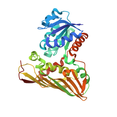
Structural insights into the catalytic and substrate recognition mechanisms of bacterial l-arabinose 1-dehydrogenase.

Watanabe, Y.Iga, C.Watanabe, Y.Watanabe, S.

(2019) FEBS Lett 593: 1257-1266

  • DOI: https://doi.org/10.1002/1873-3468.13424
  • Primary Citation of Related Structures:  
    6JNJ, 6JNK

  • PubMed Abstract: 

    In Azospirillum brasilense, a gram-negative nitrogen-fixing bacterium, l-arabinose is converted to α-ketoglutarate through a nonphosphorylative metabolic pathway. In the first step in the pathway, l-arabinose is oxidized to l-arabino-γ-lactone by NAD(P)-dependent l-arabinose 1-dehydrogenase (AraDH) belonging to the glucose-fructose oxidoreductase/inositol dehydrogenase/rhizopine catabolism protein (Gfo/Idh/MocA) family. Here, we determined the crystal structures of apo- and NADP-bound AraDH at 1.5 and 2.2 Å resolutions, respectively. A docking model of l-arabinose and NADP-bound AraDH and structure-based mutational analyses suggest that Lys91 or Asp169 serves as a catalytic base and that Glu147, His153, and Asn173 are responsible for substrate recognition. In particular, Asn173 may play a role in the discrimination between l-arabinose and d-xylose, the C4 epimer of l-arabinose.


  • Organizational Affiliation

    Department of Bioscience, Graduate School of Agriculture, Ehime University, Matsuyama, Japan.


Macromolecules
Find similar proteins by:  (by identity cutoff)  |  3D Structure
Entity ID: 1
MoleculeChains Sequence LengthOrganismDetailsImage
L-arabinose 1-dehydrogenase (NAD(P)(+))
A, B, C, D
310Azospirillum brasilenseMutation(s): 0 
Gene Names: araA
EC: 1.1.1.376 (PDB Primary Data), 1.1.1.120 (PDB Primary Data), 1.1.1.48 (PDB Primary Data)
UniProt
Find proteins for Q53TZ2 (Azospirillum brasilense)
Explore Q53TZ2 
Go to UniProtKB:  Q53TZ2
Entity Groups  
Sequence Clusters30% Identity50% Identity70% Identity90% Identity95% Identity100% Identity
UniProt GroupQ53TZ2
Sequence Annotations
Expand
  • Reference Sequence
Experimental Data & Validation

Experimental Data

  • Method: X-RAY DIFFRACTION
  • Resolution: 2.20 Å
  • R-Value Free: 0.268 
  • R-Value Work: 0.209 
  • R-Value Observed: 0.212 
  • Space Group: P 1 21 1
Unit Cell:
Length ( Å )Angle ( ˚ )
a = 70.745α = 90
b = 97.526β = 104.15
c = 104.762γ = 90
Software Package:
Software NamePurpose
PHENIXrefinement
XDSdata scaling
PDB_EXTRACTdata extraction
PHASERphasing

Structure Validation

View Full Validation Report


Ligand Structure Quality Assessment 


Entry History 

Deposition Data

Revision History  (Full details and data files)

  • Version 1.0: 2019-05-15
    Type: Initial release
  • Version 1.1: 2019-05-22
    Changes: Data collection, Database references
  • Version 1.2: 2019-06-26
    Changes: Data collection, Database references, Source and taxonomy
  • Version 1.3: 2024-03-27
    Changes: Data collection, Database references