6JKA | pdb_00006jka

Crystal structure of metallo-beta-lactamse, IMP-1, in complex with a thiazole-bearing inhibitor


Experimental Data Snapshot

  • Method: X-RAY DIFFRACTION
  • Resolution: 2.01 Å
  • R-Value Free: 
    0.250 (Depositor), 0.250 (DCC) 
  • R-Value Work: 
    0.193 (Depositor), 0.194 (DCC) 
  • R-Value Observed: 
    0.196 (Depositor) 

Starting Model: experimental
View more details

wwPDB Validation 3D Report Full Report

Validation slider image for 6JKA

Ligand Structure Quality Assessment 


This is version 1.2 of the entry. See complete history

Literature

Identification of the Inhibitory Compounds for Metallo-beta-lactamases and Structural Analysis of the Binding Modes.

Kamo, T.Kuroda, K.Kondo, S.Hayashi, U.Fudo, S.Yoneda, T.Takaya, A.Nukaga, M.Hoshino, T.

(2021) Chem Pharm Bull (Tokyo) 69: 1179-1183

  • DOI: https://doi.org/10.1248/cpb.c21-00611
  • Primary Citation Related Structures: 
    6JKA, 6JKB

  • PubMed Abstract: 

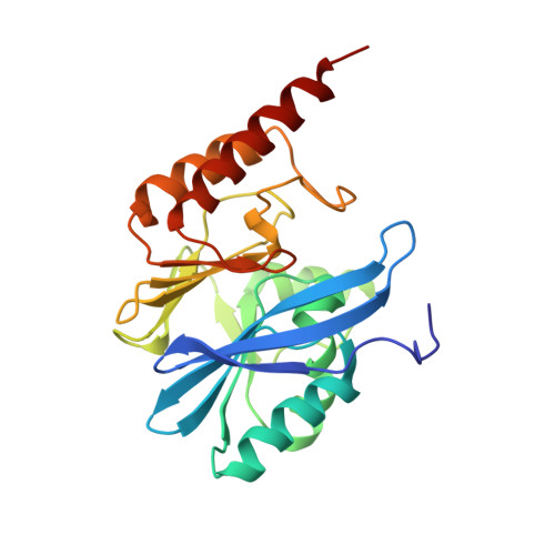
    Metallo-β-lactamases (MBLs) are significant threats to humans because they deteriorate many kinds of β-lactam antibiotics and are key enzymes responsible for multi-drug resistance of bacterial pathogens. As a result of in vitro screening, two compounds were identified as potent inhibitors of two kinds of MBLs: imipenemase (IMP-1) and New Delhi metallo-β-lactamase (NDM-1). The binding structure of one of the identified compounds was clarified by an X-ray crystal analysis in complex with IMP-1, in which two possible binding poses were observed. Molecular dynamics (MD) simulations were performed by building two calculation models from the respective binding poses. The compound was stably bound to the catalytic site during the simulation in one pose. The binding model between NDM-1 and the compound was constructed for MD simulation. Calculation results for NDM-1 were similar to those of IMP-1. The simulation suggested that the binding of the identified inhibitory compound was also durable in the catalytic site of NDM-1. The compound will be a sound basis for the development of the inhibitors for MBLs.


  • Organizational Affiliation
    • Graduate School of Pharmaceutical Sciences, Chiba University.

Macromolecule Content 

  • Total Structure Weight: 101.61 kDa 
  • Atom Count: 7,419 
  • Modeled Residue Count: 895 
  • Deposited Residue Count: 912 
  • Unique protein chains: 1

Macromolecules

Find similar proteins by:|  3D Structure
Entity ID: 1
MoleculeChains  Sequence LengthOrganismDetailsImage
Metallo-beta-lactamase type 2
A, B, C, D
228Serratia marcescensMutation(s): 0 
EC: 3.5.2.6 (PDB Primary Data), 3.1.26 (UniProt)
UniProt
Find proteins for P52699 (Serratia marcescens)
Explore P52699 
Go to UniProtKB:  P52699
Entity Groups
Sequence Clusters30% Identity50% Identity70% Identity90% Identity95% Identity100% Identity
UniProt GroupP52699
Sequence Annotations
Expand
Reference Sequence

Small Molecules

Ligands 3 Unique
IDChains Name / Formula / InChI Key2D Diagram3D Interactions
BQU

Query on BQU


Download:Ideal Coordinates CCD File
J [auth B]3-[2-azanyl-5-[2-(phenoxymethyl)-1,3-thiazol-4-yl]phenyl]propanoic acid
C19 H18 N2 O3 S
RXPAQUQGUPYCSG-UHFFFAOYSA-N
BS0

Query on BS0


Download:Ideal Coordinates CCD File
G [auth A],
M [auth C]
6-[2-(phenoxymethyl)-1,3-thiazol-4-yl]-3,4-dihydro-1H-quinolin-2-one
C19 H16 N2 O2 S
QXHYTMRDSBLWLI-UHFFFAOYSA-N
ZN

Query on ZN


Download:Ideal Coordinates CCD File
E [auth A]
F [auth A]
H [auth B]
I [auth B]
K [auth C]
E [auth A],
F [auth A],
H [auth B],
I [auth B],
K [auth C],
L [auth C],
N [auth D],
O [auth D]
ZINC ION
Zn
PTFCDOFLOPIGGS-UHFFFAOYSA-N

Experimental Data & Validation

Experimental Data

  • Method: X-RAY DIFFRACTION
  • Resolution: 2.01 Å
  • R-Value Free:  0.250 (Depositor), 0.250 (DCC) 
  • R-Value Work:  0.193 (Depositor), 0.194 (DCC) 
  • R-Value Observed: 0.196 (Depositor) 
Space Group: P 21 21 21
Unit Cell:
Length ( Å )Angle ( ˚ )
a = 48.477α = 90
b = 77.764β = 90
c = 261.527γ = 90
Software Package:
Software NamePurpose
PHENIXrefinement
SCALEPACKdata scaling
PDB_EXTRACTdata extraction
HKL-2000data reduction
MOLREPphasing

Structure Validation

View Full Validation Report


Ligand Structure Quality Assessment 


Entry History 

Deposition Data

Revision History  (Full details and data files)

  • Version 1.0: 2020-03-25
    Type: Initial release
  • Version 1.1: 2021-12-15
    Changes: Database references, Derived calculations
  • Version 1.2: 2023-11-22
    Changes: Data collection, Refinement description