6JI8

Structure of RyR2 (F/apoCaM dataset)


Experimental Data Snapshot

  • Method: ELECTRON MICROSCOPY
  • Resolution: 3.60 Å
  • Aggregation State: PARTICLE 
  • Reconstruction Method: SINGLE PARTICLE 

wwPDB Validation   3D Report Full Report


This is version 1.2 of the entry. See complete history


Literature

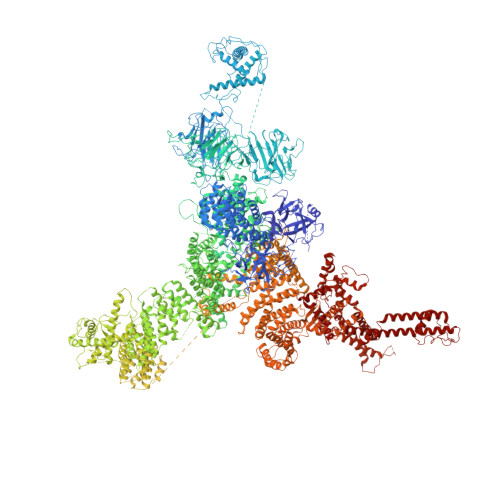
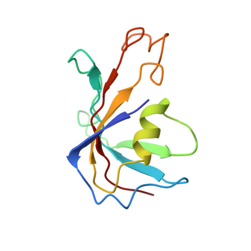
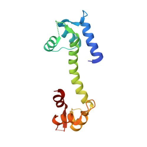
Modulation of cardiac ryanodine receptor 2 by calmodulin.

Gong, D.Chi, X.Wei, J.Zhou, G.Huang, G.Zhang, L.Wang, R.Lei, J.Chen, S.R.W.Yan, N.

(2019) Nature 572: 347-351

  • DOI: https://doi.org/10.1038/s41586-019-1377-y
  • Primary Citation of Related Structures:  
    6JI0, 6JI8, 6JII, 6JIU, 6JIY, 6JRR, 6JRS, 6JV2

  • PubMed Abstract: 
  • The high-conductance intracellular calcium (Ca 2+ ) channel RyR2 is essential for the coupling of excitation and contraction in cardiac muscle. Among various modulators, calmodulin (CaM) regulates RyR2 in a Ca 2+ -dependent manner. Here we reveal the regulatory mechanism by which porcine RyR2 is modulated by human CaM through the structural determination of RyR2 under eight conditions ...

    The high-conductance intracellular calcium (Ca 2+ ) channel RyR2 is essential for the coupling of excitation and contraction in cardiac muscle. Among various modulators, calmodulin (CaM) regulates RyR2 in a Ca 2+ -dependent manner. Here we reveal the regulatory mechanism by which porcine RyR2 is modulated by human CaM through the structural determination of RyR2 under eight conditions. Apo-CaM and Ca 2+ -CaM bind to distinct but overlapping sites in an elongated cleft formed by the handle, helical and central domains. The shift in CaM-binding sites on RyR2 is controlled by Ca 2+ binding to CaM, rather than to RyR2. Ca 2+ -CaM induces rotations and intradomain shifts of individual central domains, resulting in pore closure of the PCB95 and Ca 2+ -activated channel. By contrast, the pore of the ATP, caffeine and Ca 2+ -activated channel remains open in the presence of Ca 2+ -CaM, which suggests that Ca 2+ -CaM is one of the many competing modulators of RyR2 gating.


    Organizational Affiliation

    Department of Molecular Biology, Princeton University, Princeton, NJ, USA. nyan@princeton.edu.


Macromolecules
Find similar proteins by:  (by identity cutoff)  |  3D Structure
Entity ID: 1
MoleculeChainsSequence LengthOrganismDetailsImage
RyR2
A, D, G, J
4,968Sus scrofaMutation(s): 0 
Entity Groups  
Sequence Clusters30% Identity50% Identity70% Identity90% Identity95% Identity100% Identity
Protein Feature View
Expand
  • Reference Sequence
Find similar proteins by:  (by identity cutoff)  |  3D Structure
Entity ID: 2
MoleculeChainsSequence LengthOrganismDetailsImage
Peptidyl-prolyl cis-trans isomerase FKBP1B
B, E, H, K
108Homo sapiensMutation(s): 0 
Gene Names: FKBP1BFKBP12.6FKBP1LFKBP9OTK4
EC: 5.2.1.8
UniProt & NIH Common Fund Data Resources
Find proteins for P68106 (Homo sapiens)
Explore P68106 
Go to UniProtKB:  P68106
PHAROS:  P68106
Entity Groups  
Sequence Clusters30% Identity50% Identity70% Identity90% Identity95% Identity100% Identity
UniProt GroupP68106
Protein Feature View
Expand
  • Reference Sequence
Find similar proteins by:  (by identity cutoff)  |  3D Structure
Entity ID: 3
MoleculeChainsSequence LengthOrganismDetailsImage
Calmodulin-1
C, F, I, L
149Homo sapiensMutation(s): 0 
Gene Names: CALM1CALMCAMCAM1
UniProt & NIH Common Fund Data Resources
Find proteins for P0DP23 (Homo sapiens)
Explore P0DP23 
Go to UniProtKB:  P0DP23
PHAROS:  P0DP23
Entity Groups  
Sequence Clusters30% Identity50% Identity70% Identity90% Identity95% Identity100% Identity
UniProt GroupP0DP23
Protein Feature View
Expand
  • Reference Sequence
Small Molecules
Experimental Data & Validation

Experimental Data

  • Method: ELECTRON MICROSCOPY
  • Resolution: 3.60 Å
  • Aggregation State: PARTICLE 
  • Reconstruction Method: SINGLE PARTICLE 
EM Software:
TaskSoftware PackageVersion
RECONSTRUCTIONRELION2.0

Structure Validation

View Full Validation Report


Entry History 

Deposition Data

Revision History  (Full details and data files)

  • Version 1.0: 2019-07-17
    Type: Initial release
  • Version 1.1: 2019-08-28
    Changes: Data collection, Database references, Source and taxonomy, Structure summary
  • Version 1.2: 2019-11-06
    Changes: Data collection, Other