6JFZ

GluK3 receptor complex with UBP310


Experimental Data Snapshot

  • Method: ELECTRON MICROSCOPY
  • Resolution: 7.60 Å
  • Aggregation State: PARTICLE 
  • Reconstruction Method: SINGLE PARTICLE 

wwPDB Validation   3D Report Full Report


This is version 1.2 of the entry. See complete history


Literature

Structural and Functional Insights into GluK3-kainate Receptor Desensitization and Recovery.

Kumari, J.Vinnakota, R.Kumar, J.

(2019) Sci Rep 9: 10254-10254

  • DOI: https://doi.org/10.1038/s41598-019-46770-z
  • Primary Citation of Related Structures:  
    6JFY, 6JFZ, 6JMV

  • PubMed Abstract: 

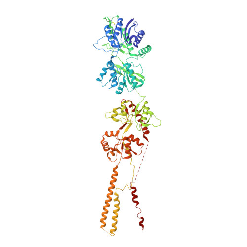
    GluK3-kainate receptors are atypical members of the iGluR family that reside at both the pre- and postsynapse and play a vital role in the regulation of synaptic transmission. For a better understanding of structural changes that underlie receptor functions, GluK3 receptors were trapped in desensitized and resting/closed states and structures analyzed using single particle cryo-electron microscopy. While the desensitized GluK3 has domain organization as seen earlier for another kainate receptor-GluK2, antagonist bound GluK3 trapped a resting state with only two LBD domains in dimeric arrangement necessary for receptor activation. Using structures as a guide, we show that the N-linked glycans at the interface of GluK3 ATD and LBD likely mediate inter-domain interactions and attune receptor-gating properties. The mutational analysis also identified putative N-glycan interacting residues. Our results provide a molecular framework for understanding gating properties unique to GluK3 and exploring the role of N-linked glycosylation in their modulation.


  • Organizational Affiliation

    Laboratory of Membrane Protein Biology, National Centre for Cell Science, NCCS Complex, S. P. Pune University, Maharashtra, Pune, 411007, India.


Macromolecules
Find similar proteins by:  (by identity cutoff)  |  3D Structure
Entity ID: 1
MoleculeChains Sequence LengthOrganismDetailsImage
Glutamate receptor ionotropic, kainate 3A,
B,
C [auth D],
D [auth C]
809Rattus norvegicusMutation(s): 3 
Gene Names: Grik3Glur7
Membrane Entity: Yes 
UniProt
Find proteins for P42264 (Rattus norvegicus)
Explore P42264 
Go to UniProtKB:  P42264
Entity Groups  
Sequence Clusters30% Identity50% Identity70% Identity90% Identity95% Identity100% Identity
UniProt GroupP42264
Sequence Annotations
Expand
  • Reference Sequence
Experimental Data & Validation

Experimental Data

  • Method: ELECTRON MICROSCOPY
  • Resolution: 7.60 Å
  • Aggregation State: PARTICLE 
  • Reconstruction Method: SINGLE PARTICLE 
EM Software:
TaskSoftware PackageVersion
MODEL REFINEMENTPHENIX1.10.1-2155
RECONSTRUCTIONcryoSPARC2

Structure Validation

View Full Validation Report


Entry History & Funding Information

Deposition Data


Funding OrganizationLocationGrant Number
Wellcome TrustIndiaIA/I/13/2/501023
Department of Biotechnology (India)IndiaDBT/PR12422/MED/31/287/2014

Revision History  (Full details and data files)

  • Version 1.0: 2019-07-24
    Type: Initial release
  • Version 1.1: 2019-07-31
    Changes: Data collection, Database references
  • Version 1.2: 2019-11-06
    Changes: Data collection, Other