


K2U bound crystal peptide deformylase from Acinetobacter baumanii
Lee, I.H., Ho, T.H., Kang, L.W.To be published.
Experimental Data Snapshot
Starting Model: experimental
View more details
Entity ID: 1 | |||||
|---|---|---|---|---|---|
| Molecule | Chains | Sequence Length | Organism | Details | Image |
| Peptide deformylase | 171 | Acinetobacter baumannii MRSN 3527 | Mutation(s): 0 Gene Names: def_1, def, T630_0214 EC: 3.5.1.88 | 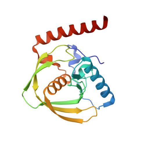 | |
UniProt | |||||
Entity Groups | |||||
| Sequence Clusters | 30% Identity50% Identity70% Identity90% Identity95% Identity100% Identity | ||||
| UniProt Group | B0VNL8 | ||||
Sequence AnnotationsExpand | |||||
Reference Sequence | |||||
| Ligands 2 Unique | |||||
|---|---|---|---|---|---|
| ID | Chains | Name / Formula / InChI Key | 2D Diagram | 3D Interactions | |
| K2U Download:Ideal Coordinates CCD File | D [auth A], F [auth B] | (3~{R},4~{R})-4-oxidanyl-3-(phenylmethyl)-4-(phenylmethylsulfanyl)butanoic acid C18 H20 O3 S OBXQOECJZPGJTO-SJLPKXTDSA-N |  | ||
| ZN Download:Ideal Coordinates CCD File | C [auth A], E [auth B] | ZINC ION Zn PTFCDOFLOPIGGS-UHFFFAOYSA-N |  | ||
| Length ( Å ) | Angle ( ˚ ) |
|---|---|
| a = 40.062 | α = 90 |
| b = 40.062 | β = 90 |
| c = 188.271 | γ = 120 |
| Software Name | Purpose |
|---|---|
| REFMAC | refinement |
| HKL-2000 | data scaling |
| HKL-2000 | data collection |
| HKL-2000 | data reduction |
| MOLREP | phasing |


