The role of gelsolin domain 3 in familial amyloidosis (Finnish type).
Zorgati, H., Larsson, M., Ren, W., Sim, A.Y.L., Gettemans, J., Grimes, J.M., Li, W., Robinson, R.C.(2019) Proc Natl Acad Sci U S A 116: 13958-13963
- PubMed: 31243148 Search on PubMedSearch on PubMed Central
- DOI: https://doi.org/10.1073/pnas.1902189116
- Primary Citation Related Structures:
6JCO, 6JEG, 6JEH - PubMed Abstract:
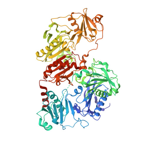
In the disease familial amyloidosis, Finnish type (FAF), also known as AGel amyloidosis (AGel), the mechanism by which point mutations in the calcium-regulated actin-severing protein gelsolin lead to furin cleavage is not understood in the intact protein. Here, we provide a structural and biochemical characterization of the FAF variants. X-ray crystallography structures of the FAF mutant gelsolins demonstrate that the mutations do not significantly disrupt the calcium-free conformations of gelsolin. Small-angle X-ray-scattering (SAXS) studies indicate that the FAF calcium-binding site mutants are slower to activate, whereas G167R is as efficient as the wild type. Actin-regulating studies of the gelsolins at the furin cleavage pH (6.5) show that the mutant gelsolins are functional, suggesting that they also adopt relatively normal active conformations. Deletion of gelsolin domains leads to sensitization to furin cleavage, and nanobody-binding protects against furin cleavage. These data indicate instability in the second domain of gelsolin (G2), since loss or gain of G2-stabilizing interactions impacts the efficiency of cleavage by furin. To demonstrate this principle, we engineered non-FAF mutations in G3 that disrupt the G2-G3 interface in the calcium-activated structure. These mutants led to increased furin cleavage. We carried out molecular dynamics (MD) simulations on the FAF and non-FAF mutant G2-G3 fragments of gelsolin. All mutants showed an increase in the distance between the center of masses of the 2 domains (G2 and G3). Since G3 covers the furin cleavage site on G2 in calcium-activated gelsolin, this suggests that destabilization of this interface is a critical step in cleavage.
- Institute of Molecular and Cell Biology, Agency for Science, Technology and Research (A*STAR), 138673 Singapore, Singapore.
Organizational Affiliation: