6JBX

Crystal structure of Streptococcus pneumoniae FabT in complex with DNA


Experimental Data Snapshot

  • Method: X-RAY DIFFRACTION
  • Resolution: 2.20 Å
  • R-Value Free: 0.221 
  • R-Value Work: 0.188 
  • R-Value Observed: 0.189 

wwPDB Validation   3D Report Full Report


Ligand Structure Quality Assessment 


This is version 1.2 of the entry. See complete history


Literature

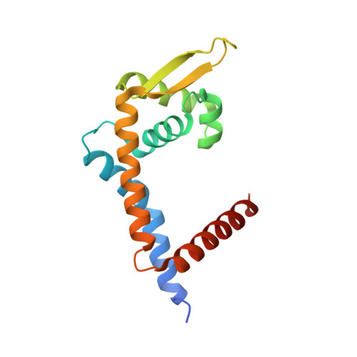
Structural insights into repression of the Pneumococcal fatty acid synthesis pathway by repressor FabT and co-repressor acyl-ACP.

Zuo, G.Chen, Z.P.Jiang, Y.L.Zhu, Z.Ding, C.Zhang, Z.Chen, Y.Zhou, C.Z.Li, Q.

(2019) FEBS Lett 593: 2730-2741

  • DOI: https://doi.org/10.1002/1873-3468.13534
  • Primary Citation of Related Structures:  
    6JBX

  • PubMed Abstract: 

    The Streptococcus pneumoniae fatty acid synthesis (FAS) pathway is globally controlled at the transcriptional level by the repressor FabT and its co-repressor acyl carrier protein (acyl-ACP), the intermediate of phospholipid synthesis. Here, we report the crystal structure of FabT complexed with a 23-bp dsDNA, which indicates that FabT is a weak repressor with low DNA-binding affinity in the absence of acyl-ACP. Modification of ACP with a long-chain fatty acid is necessary for the formation of a stable complex with FabT, mimicked in vitro by cross-linking, which significantly elevates the DNA-binding affinity of FabT. Altogether, we propose a putative working model of gene repression under the double control of FabT and acyl-ACP, elucidating a distinct repression network for Pneumococcus to precisely coordinate FAS.


  • Organizational Affiliation

    Hefei National Laboratory for Physical Sciences at the Microscale and School of Life Sciences, University of Science and Technology of China, Hefei, Anhui, China.


Macromolecules

Find similar proteins by:  (by identity cutoff)  |  3D Structure
Entity ID: 1
MoleculeChains Sequence LengthOrganismDetailsImage
Fatty acid biosynthesis transcriptional regulator
A, B
152Streptococcus pneumoniaeMutation(s): 0 
Gene Names: marR_1fabTmarR
UniProt
Find proteins for Q8DR18 (Streptococcus pneumoniae (strain ATCC BAA-255 / R6))
Explore Q8DR18 
Go to UniProtKB:  Q8DR18
Entity Groups  
Sequence Clusters30% Identity50% Identity70% Identity90% Identity95% Identity100% Identity
UniProt GroupQ8DR18
Sequence Annotations
Expand
  • Reference Sequence

Find similar nucleic acids by:  Sequence   |   3D Structure  

Entity ID: 2
MoleculeChains LengthOrganismImage
DNA (5'-D(*AP*AP*TP*AP*GP*TP*TP*TP*GP*AP*CP*TP*GP*TP*CP*AP*AP*AP*TP*TP*AP*TP*G)-3')23Streptococcus pneumoniae
Sequence Annotations
Expand
  • Reference Sequence

Find similar nucleic acids by:  Sequence   |   3D Structure  

Entity ID: 3
MoleculeChains LengthOrganismImage
DNA (5'-D(*CP*AP*TP*AP*AP*TP*TP*TP*GP*AP*CP*AP*GP*TP*CP*AP*AP*AP*CP*TP*AP*TP*T)-3')23Streptococcus pneumoniae
Sequence Annotations
Expand
  • Reference Sequence
Experimental Data & Validation

Experimental Data

  • Method: X-RAY DIFFRACTION
  • Resolution: 2.20 Å
  • R-Value Free: 0.221 
  • R-Value Work: 0.188 
  • R-Value Observed: 0.189 
  • Space Group: P 1 21 1
Unit Cell:
Length ( Å )Angle ( ˚ )
a = 73.022α = 90
b = 60.868β = 105.4
c = 74.086γ = 90
Software Package:
Software NamePurpose
REFMACrefinement
HKL-2000data reduction
HKL-2000data scaling
PHENIXmodel building

Structure Validation

View Full Validation Report


Ligand Structure Quality Assessment 


Entry History & Funding Information

Deposition Data


Funding OrganizationLocationGrant Number
National Natural Science Foundation of ChinaChina31570728

Revision History  (Full details and data files)

  • Version 1.0: 2019-07-24
    Type: Initial release
  • Version 1.1: 2019-10-23
    Changes: Data collection, Database references
  • Version 1.2: 2024-03-27
    Changes: Data collection, Database references