6JBD

Phosphotransferase-ATP complex related to CoA biosynthesis pathway


Experimental Data Snapshot

  • Method: X-RAY DIFFRACTION
  • Resolution: 2.50 Å
  • R-Value Free: 0.259 
  • R-Value Work: 0.219 

wwPDB Validation   3D Report Full Report


Ligand Structure Quality Assessment 


This is version 1.2 of the entry. See complete history


Literature

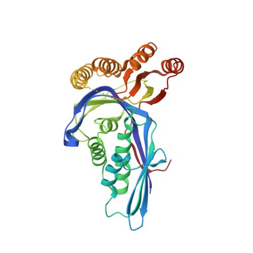
Crystal structure of pantoate kinase from Thermococcus kodakarensis.

Kita, A.Kishimoto, A.Shimosaka, T.Tomita, H.Yokooji, Y.Imanaka, T.Atomi, H.Miki, K.

(2020) Proteins 88: 718-724

  • DOI: https://doi.org/10.1002/prot.25852
  • Primary Citation of Related Structures:  
    6JBC, 6JBD

  • PubMed Abstract: 

    The coenzyme A biosynthesis pathways in most archaea involve two unique enzymes, pantoate kinase and phosphopantothenate synthetase, to convert pantoate to 4'-phosphopantothenate. Here, we report the first crystal structure of pantoate kinase from the hyperthermophilic archaeon, Thermococcus kodakarensis and its complex with ATP and a magnesium ion. The electron density for the adenosine moiety of ATP was very weak, which most likely relates to its broad nucleotide specificity. Based on the structure of the active site that contains a glycerol molecule, the pantoate binding site and the roles of the highly conserved residues are suggested.


  • Organizational Affiliation

    Institute for Integrated Radiation and Nuclear Science, Kyoto University, Osaka, Japan.


Macromolecules
Find similar proteins by:  (by identity cutoff)  |  3D Structure
Entity ID: 1
MoleculeChains Sequence LengthOrganismDetailsImage
Pantoate kinase300Thermococcus kodakarensis KOD1Mutation(s): 0 
Gene Names: TK2141
EC: 2.7.1.169
UniProt
Find proteins for Q5JHF1 (Thermococcus kodakarensis (strain ATCC BAA-918 / JCM 12380 / KOD1))
Explore Q5JHF1 
Go to UniProtKB:  Q5JHF1
Entity Groups  
Sequence Clusters30% Identity50% Identity70% Identity90% Identity95% Identity100% Identity
UniProt GroupQ5JHF1
Sequence Annotations
Expand
  • Reference Sequence
Small Molecules
Ligands 7 Unique
IDChains Name / Formula / InChI Key2D Diagram3D Interactions
ATP
Query on ATP

Download Ideal Coordinates CCD File 
G [auth A]ADENOSINE-5'-TRIPHOSPHATE
C10 H16 N5 O13 P3
ZKHQWZAMYRWXGA-KQYNXXCUSA-N
PGE
Query on PGE

Download Ideal Coordinates CCD File 
F [auth A]TRIETHYLENE GLYCOL
C6 H14 O4
ZIBGPFATKBEMQZ-UHFFFAOYSA-N
GOL
Query on GOL

Download Ideal Coordinates CCD File 
B [auth A],
C [auth A],
D [auth A]
GLYCEROL
C3 H8 O3
PEDCQBHIVMGVHV-UHFFFAOYSA-N
EDO
Query on EDO

Download Ideal Coordinates CCD File 
E [auth A]1,2-ETHANEDIOL
C2 H6 O2
LYCAIKOWRPUZTN-UHFFFAOYSA-N
CA
Query on CA

Download Ideal Coordinates CCD File 
I [auth A],
J [auth A],
K [auth A]
CALCIUM ION
Ca
BHPQYMZQTOCNFJ-UHFFFAOYSA-N
MG
Query on MG

Download Ideal Coordinates CCD File 
H [auth A]MAGNESIUM ION
Mg
JLVVSXFLKOJNIY-UHFFFAOYSA-N
NA
Query on NA

Download Ideal Coordinates CCD File 
L [auth A],
M [auth A]
SODIUM ION
Na
FKNQFGJONOIPTF-UHFFFAOYSA-N
Experimental Data & Validation

Experimental Data

  • Method: X-RAY DIFFRACTION
  • Resolution: 2.50 Å
  • R-Value Free: 0.259 
  • R-Value Work: 0.219 
  • Space Group: P 3 2 1
Unit Cell:
Length ( Å )Angle ( ˚ )
a = 95.323α = 90
b = 95.323β = 90
c = 60.691γ = 120
Software Package:
Software NamePurpose
SCALEPACKdata scaling
CNSrefinement
PDB_EXTRACTdata extraction
HKL-2000data reduction
SHARPphasing

Structure Validation

View Full Validation Report


Ligand Structure Quality Assessment 


Entry History & Funding Information

Deposition Data


Funding OrganizationLocationGrant Number
Japan Society for the Promotion of ScienceJapan24.2716
Japan Society for the Promotion of ScienceJapanJP17J08246
Japan Society for the Promotion of ScienceJapanJP18H03934
Other privateJapan--

Revision History  (Full details and data files)

  • Version 1.0: 2020-01-29
    Type: Initial release
  • Version 1.1: 2020-08-19
    Changes: Database references, Derived calculations
  • Version 1.2: 2024-03-27
    Changes: Data collection, Database references