6J8M | pdb_00006j8m

Low-dose structure of bovine heart cytochrome c oxidase in the fully oxidized state determined using 30 keV X-ray


Experimental Data Snapshot

  • Method: X-RAY DIFFRACTION
  • Resolution: 1.90 Å
  • R-Value Free: 
    0.199 (Depositor), 0.225 (DCC) 
  • R-Value Work: 
    0.172 (Depositor), 0.175 (DCC) 
  • R-Value Observed: 
    0.173 (Depositor) 

Starting Model: experimental
View more details

wwPDB Validation 3D Report Full Report

Validation slider image for 6J8M

Ligand Structure Quality Assessment 


This is version 1.3 of the entry. See complete history

Literature

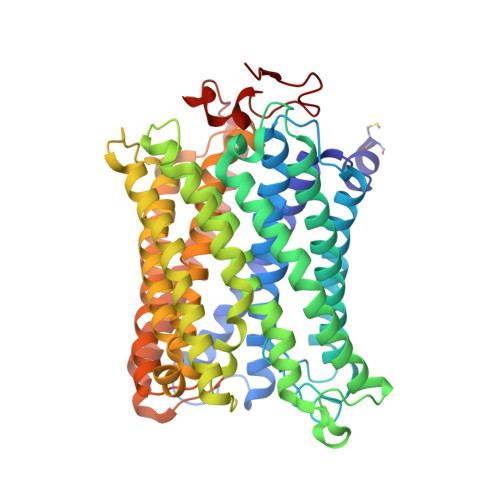
Low-dose X-ray structure analysis of cytochrome c oxidase utilizing high-energy X-rays.

Ueno, G.Shimada, A.Yamashita, E.Hasegawa, K.Kumasaka, T.Shinzawa-Itoh, K.Yoshikawa, S.Tsukihara, T.Yamamoto, M.

(2019) J Synchrotron Radiat 26: 912-921

  • DOI: https://doi.org/10.1107/S1600577519006805
  • Primary Citation Related Structures: 
    6J8M

  • PubMed Abstract: 

    To investigate the effect of high-energy X-rays on site-specific radiation-damage, low-dose diffraction data were collected from radiation-sensitive crystals of the metal enzyme cytochrome c oxidase. Data were collected at the Structural Biology I beamline (BL41XU) at SPring-8, using 30 keV X-rays and a highly sensitive pixel array detector equipped with a cadmium telluride sensor. The experimental setup of continuous sample translation using multiple crystals allowed the average diffraction weighted dose per data set to be reduced to 58 kGy, and the resulting data revealed a ligand structure featuring an identical bond length to that in the damage-free structure determined using an X-ray free-electron laser. However, precise analysis of the residual density around the ligand structure refined with the synchrotron data showed the possibility of a small level of specific damage, which might have resulted from the accumulated dose of 58 kGy per data set. Further investigation of the photon-energy dependence of specific damage, as assessed by variations in UV-vis absorption spectra, was conducted using an on-line spectrometer at various energies ranging from 10 to 30 keV. No evidence was found for specific radiation damage being energy dependent.


  • Organizational Affiliation
    • SR Life Science Instrumentation Team, Life Science Research Infrastructure Group, Advanced Photon Technology Division, RIKEN SPring-8 Center, 1-1-1 Kouto, Sayo-cho, Sayo-gun, Hyogo 679-5148, Japan.

Macromolecule Content 

  • Total Structure Weight: 442.87 kDa 
  • Atom Count: 33,893 
  • Modeled Residue Count: 3,558 
  • Deposited Residue Count: 3,614 
  • Unique protein chains: 13

Macromolecules

Find similar proteins by:|  3D Structure
Entity ID: 1
MoleculeChains  Sequence LengthOrganismDetailsImage
Cytochrome c oxidase subunit 1
A, N
514Bos taurusMutation(s): 0 
EC: 1.9.3.1 (PDB Primary Data), 7.1.1.9 (UniProt)
Membrane Entity: Yes 
UniProt
Find proteins for P00396 (Bos taurus)
Explore P00396 
Go to UniProtKB:  P00396
Entity Groups
Sequence Clusters30% Identity50% Identity70% Identity90% Identity95% Identity100% Identity
UniProt GroupP00396
Sequence Annotations
Expand
Reference Sequence
Find similar proteins by:|  3D Structure
Entity ID: 2
MoleculeChains  Sequence LengthOrganismDetailsImage
Cytochrome c oxidase subunit 2
B, O
227Bos taurusMutation(s): 0 
EC: 7.1.1.9
Membrane Entity: Yes 
UniProt
Find proteins for P68530 (Bos taurus)
Explore P68530 
Go to UniProtKB:  P68530
Entity Groups
Sequence Clusters30% Identity50% Identity70% Identity90% Identity95% Identity100% Identity
UniProt GroupP68530
Sequence Annotations
Expand
Reference Sequence
Find similar proteins by:|  3D Structure
Entity ID: 3
MoleculeChains  Sequence LengthOrganismDetailsImage
Cytochrome c oxidase subunit 3
C, P
261Bos taurusMutation(s): 0 
EC: 7.1.1.9
Membrane Entity: Yes 
UniProt
Find proteins for P00415 (Bos taurus)
Explore P00415 
Go to UniProtKB:  P00415
Entity Groups
Sequence Clusters30% Identity50% Identity70% Identity90% Identity95% Identity100% Identity
UniProt GroupP00415
Sequence Annotations
Expand
Reference Sequence
Find similar proteins by:|  3D Structure
Entity ID: 4
MoleculeChains  Sequence LengthOrganismDetailsImage
Cytochrome c oxidase subunit 4 isoform 1
D, Q
147Bos taurusMutation(s): 0 
Membrane Entity: Yes 
UniProt
Find proteins for P00423 (Bos taurus)
Explore P00423 
Go to UniProtKB:  P00423
Entity Groups
Sequence Clusters30% Identity50% Identity70% Identity90% Identity95% Identity100% Identity
UniProt GroupP00423
Sequence Annotations
Expand
Reference Sequence
Find similar proteins by:|  3D Structure
Entity ID: 5
MoleculeChains  Sequence LengthOrganismDetailsImage
Cytochrome c oxidase subunit 5A
E, R
109Bos taurusMutation(s): 0 
Membrane Entity: Yes 
UniProt
Find proteins for P00426 (Bos taurus)
Explore P00426 
Go to UniProtKB:  P00426
Entity Groups
Sequence Clusters30% Identity50% Identity70% Identity90% Identity95% Identity100% Identity
UniProt GroupP00426
Sequence Annotations
Expand
Reference Sequence
Find similar proteins by:|  3D Structure
Entity ID: 6
MoleculeChains  Sequence LengthOrganismDetailsImage
Cytochrome c oxidase subunit 5B
F, S
98Bos taurusMutation(s): 0 
Membrane Entity: Yes 
UniProt
Find proteins for P00428 (Bos taurus)
Explore P00428 
Go to UniProtKB:  P00428
Entity Groups
Sequence Clusters30% Identity50% Identity70% Identity90% Identity95% Identity100% Identity
UniProt GroupP00428
Sequence Annotations
Expand
Reference Sequence
Find similar proteins by:|  3D Structure
Entity ID: 7
MoleculeChains  Sequence LengthOrganismDetailsImage
Cytochrome c oxidase subunit 6A2
G, T
85Bos taurusMutation(s): 0 
Membrane Entity: Yes 
UniProt
Find proteins for P07471 (Bos taurus)
Explore P07471 
Go to UniProtKB:  P07471
Entity Groups
Sequence Clusters30% Identity50% Identity70% Identity90% Identity95% Identity100% Identity
UniProt GroupP07471
Sequence Annotations
Expand
Reference Sequence
Find similar proteins by:|  3D Structure
Entity ID: 8
MoleculeChains  Sequence LengthOrganismDetailsImage
Cytochrome c oxidase subunit 6B1
H, U
85Bos taurusMutation(s): 0 
Membrane Entity: Yes 
UniProt
Find proteins for P00429 (Bos taurus)
Explore P00429 
Go to UniProtKB:  P00429
Entity Groups
Sequence Clusters30% Identity50% Identity70% Identity90% Identity95% Identity100% Identity
UniProt GroupP00429
Sequence Annotations
Expand
Reference Sequence
Find similar proteins by:|  3D Structure
Entity ID: 9
MoleculeChains  Sequence LengthOrganismDetailsImage
Cytochrome c oxidase subunit 6C
I, V
73Bos taurusMutation(s): 0 
Membrane Entity: Yes 
UniProt
Find proteins for P04038 (Bos taurus)
Explore P04038 
Go to UniProtKB:  P04038
Entity Groups
Sequence Clusters30% Identity50% Identity70% Identity90% Identity95% Identity100% Identity
UniProt GroupP04038
Sequence Annotations
Expand
Reference Sequence
Find similar proteins by:|  3D Structure
Entity ID: 10
MoleculeChains  Sequence LengthOrganismDetailsImage
Cytochrome c oxidase subunit 7A1
J, W
59Bos taurusMutation(s): 0 
Membrane Entity: Yes 
UniProt
Find proteins for P07470 (Bos taurus)
Explore P07470 
Go to UniProtKB:  P07470
Entity Groups
Sequence Clusters30% Identity50% Identity70% Identity90% Identity95% Identity100% Identity
UniProt GroupP07470
Sequence Annotations
Expand
Reference Sequence
Find similar proteins by:|  3D Structure
Entity ID: 11
MoleculeChains  Sequence LengthOrganismDetailsImage
Cytochrome c oxidase subunit 7B
K, X
56Bos taurusMutation(s): 0 
Membrane Entity: Yes 
UniProt
Find proteins for P13183 (Bos taurus)
Explore P13183 
Go to UniProtKB:  P13183
Entity Groups
Sequence Clusters30% Identity50% Identity70% Identity90% Identity95% Identity100% Identity
UniProt GroupP13183
Sequence Annotations
Expand
Reference Sequence
Find similar proteins by:|  3D Structure
Entity ID: 12
MoleculeChains  Sequence LengthOrganismDetailsImage
Cytochrome c oxidase subunit 7C
L, Y
47Bos taurusMutation(s): 0 
Membrane Entity: Yes 
UniProt
Find proteins for P00430 (Bos taurus)
Explore P00430 
Go to UniProtKB:  P00430
Entity Groups
Sequence Clusters30% Identity50% Identity70% Identity90% Identity95% Identity100% Identity
UniProt GroupP00430
Sequence Annotations
Expand
Reference Sequence
Find similar proteins by:|  3D Structure
Entity ID: 13
MoleculeChains  Sequence LengthOrganismDetailsImage
Cytochrome c oxidase subunit 8B
M, Z
46Bos taurusMutation(s): 0 
Membrane Entity: Yes 
UniProt
Find proteins for P10175 (Bos taurus)
Explore P10175 
Go to UniProtKB:  P10175
Entity Groups
Sequence Clusters30% Identity50% Identity70% Identity90% Identity95% Identity100% Identity
UniProt GroupP10175
Sequence Annotations
Expand
Reference Sequence

Small Molecules

Ligands 14 Unique
IDChains Name / Formula / InChI Key2D Diagram3D Interactions
CDL

Query on CDL


Download:Ideal Coordinates CCD File
BC [auth T],
QA [auth C],
TB [auth P],
XA [auth G]
CARDIOLIPIN
C81 H156 O17 P2
XVTUQDWPJJBEHJ-KZCWQMDCSA-L
TGL

Query on TGL


Download:Ideal Coordinates CCD File
AB [auth L]
IA [auth B]
JB [auth N]
MB [auth N]
UA [auth D]
AB [auth L],
IA [auth B],
JB [auth N],
MB [auth N],
UA [auth D],
WB [auth Q]
TRISTEAROYLGLYCEROL
C57 H110 O6
DCXXMTOCNZCJGO-UHFFFAOYSA-N
HEA

Query on HEA


Download:Ideal Coordinates CCD File
AA [auth A],
BA [auth A],
CB [auth N],
DB [auth N]
HEME-A
C49 H56 Fe N4 O6
ZGGYGTCPXNDTRV-PRYGPKJJSA-L
PEK

Query on PEK


Download:Ideal Coordinates CCD File
AC [auth T]
MA [auth C]
NA [auth C]
QB [auth P]
YA [auth G]
AC [auth T],
MA [auth C],
NA [auth C],
QB [auth P],
YA [auth G],
ZB [auth T]
(1S)-2-{[(2-AMINOETHOXY)(HYDROXY)PHOSPHORYL]OXY}-1-[(STEAROYLOXY)METHYL]ETHYL (5E,8E,11E,14E)-ICOSA-5,8,11,14-TETRAENOATE
C43 H78 N O8 P
ANRKEHNWXKCXDB-BHFWLYLHSA-N
PSC

Query on PSC


Download:Ideal Coordinates CCD File
VA [auth E],
XB [auth R]
(7R,17E,20E)-4-HYDROXY-N,N,N-TRIMETHYL-9-OXO-7-[(PALMITOYLOXY)METHYL]-3,5,8-TRIOXA-4-PHOSPHAHEXACOSA-17,20-DIEN-1-AMINIUM 4-OXIDE
C42 H81 N O8 P
JLPULHDHAOZNQI-AUSZDXHESA-O
PGV

Query on PGV


Download:Ideal Coordinates CCD File
FA [auth A]
HA [auth A]
HB [auth N]
KB [auth N]
OA [auth C]
FA [auth A],
HA [auth A],
HB [auth N],
KB [auth N],
OA [auth C],
PA [auth C],
RB [auth P],
SB [auth P]
(1R)-2-{[{[(2S)-2,3-DIHYDROXYPROPYL]OXY}(HYDROXY)PHOSPHORYL]OXY}-1-[(PALMITOYLOXY)METHYL]ETHYL (11E)-OCTADEC-11-ENOATE
C40 H77 O10 P
ADYWCMPUNIVOEA-GPJPVTGXSA-N
DMU

Query on DMU


Download:Ideal Coordinates CCD File
BB [auth M],
DC [auth Z],
TA [auth C],
VB [auth P]
DECYL-BETA-D-MALTOPYRANOSIDE
C22 H42 O11
WOQQAWHSKSSAGF-WXFJLFHKSA-N
CHD

Query on CHD


Download:Ideal Coordinates CCD File
CC [auth W]
KA [auth B]
LB [auth N]
NB [auth O]
RA [auth C]
CC [auth W],
KA [auth B],
LB [auth N],
NB [auth O],
RA [auth C],
SA [auth C],
UB [auth P],
ZA [auth J]
CHOLIC ACID
C24 H40 O5
BHQCQFFYRZLCQQ-OELDTZBJSA-N
CUA

Query on CUA


Download:Ideal Coordinates CCD File
JA [auth B],
OB [auth O]
DINUCLEAR COPPER ION
Cu2
ALKZAGKDWUSJED-UHFFFAOYSA-N
ZN

Query on ZN


Download:Ideal Coordinates CCD File
WA [auth F],
YB [auth S]
ZINC ION
Zn
PTFCDOFLOPIGGS-UHFFFAOYSA-N
CU

Query on CU


Download:Ideal Coordinates CCD File
CA [auth A],
EB [auth N]
COPPER (II) ION
Cu
JPVYNHNXODAKFH-UHFFFAOYSA-N
PER

Query on PER


Download:Ideal Coordinates CCD File
GA [auth A],
IB [auth N]
PEROXIDE ION
O2
ANAIPYUSIMHBEL-UHFFFAOYSA-N
MG

Query on MG


Download:Ideal Coordinates CCD File
DA [auth A],
FB [auth N]
MAGNESIUM ION
Mg
JLVVSXFLKOJNIY-UHFFFAOYSA-N
NA

Query on NA


Download:Ideal Coordinates CCD File
EA [auth A],
GB [auth N],
LA [auth C],
PB [auth P]
SODIUM ION
Na
FKNQFGJONOIPTF-UHFFFAOYSA-N
Modified Residues  3 Unique
IDChains TypeFormula2D DiagramParent
FME
Query on FME
A, N
L-PEPTIDE LINKINGC6 H11 N O3 SMET
TPO
Query on TPO
G, T
L-PEPTIDE LINKINGC4 H10 N O6 PTHR
SAC
Query on SAC
I, V
L-PEPTIDE LINKINGC5 H9 N O4SER

Experimental Data & Validation

Experimental Data

  • Method: X-RAY DIFFRACTION
  • Resolution: 1.90 Å
  • R-Value Free:  0.199 (Depositor), 0.225 (DCC) 
  • R-Value Work:  0.172 (Depositor), 0.175 (DCC) 
  • R-Value Observed: 0.173 (Depositor) 
Space Group: P 21 21 21
Unit Cell:
Length ( Å )Angle ( ˚ )
a = 182.02α = 90
b = 203.54β = 90
c = 177.41γ = 90
Software Package:
Software NamePurpose
XSCALEdata scaling
REFMACrefinement
PDB_EXTRACTdata extraction
XDSdata reduction
REFMACphasing

Structure Validation

View Full Validation Report


Ligand Structure Quality Assessment 


Entry History 

& Funding Information

Deposition Data


Funding OrganizationLocationGrant Number
Japan Agency for Medical Research and Development (AMED)Japan--
Japan Science and TechnologyJapan--

Revision History  (Full details and data files)

  • Version 1.0: 2019-06-26
    Type: Initial release
  • Version 1.1: 2019-07-17
    Changes: Data collection, Database references
  • Version 1.2: 2019-12-04
    Changes: Data collection
  • Version 1.3: 2023-11-22
    Changes: Data collection, Database references, Derived calculations, Refinement description, Structure summary