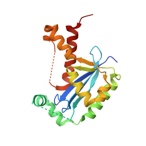
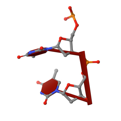
6J7Y

Human mitochondrial Oligoribonuclease in complex with DNA


Experimental Data Snapshot

  • Method: X-RAY DIFFRACTION
  • Resolution: 2.20 Å
  • R-Value Free: 0.244 
  • R-Value Work: 0.198 
  • R-Value Observed: 0.202 

Starting Model: experimental
View more details

wwPDB Validation   3D Report Full Report


This is version 1.1 of the entry. See complete history


Literature

Structural insights into nanoRNA degradation by human Rexo2.

Chu, L.Y.Agrawal, S.Chen, Y.P.Yang, W.Z.Yuan, H.S.

(2019) RNA 25: 737-746

  • DOI: https://doi.org/10.1261/rna.070557.119
  • Primary Citation of Related Structures:  
    6J7Y, 6J7Z, 6J80

  • PubMed Abstract: 

    Human RNA exoribonuclease 2 (Rexo2) is an evolutionarily conserved 3'-to-5' DEDDh-family exonuclease located primarily in mitochondria. Rexo2 degrades small RNA oligonucleotides of <5 nucleotides (nanoRNA) in a way similar to Escherichia coli Oligoribonuclease (ORN), suggesting that it plays a role in RNA turnover in mitochondria. However, how Rexo2 preferentially binds and degrades nanoRNA remains elusive. Here, we show that Rexo2 binds small RNA and DNA oligonucleotides with the highest affinity, and it is most robust in degrading small nanoRNA into mononucleotides in the presence of magnesium ions. We further determined three crystal structures of Rexo2 in complex with single-stranded RNA or DNA at resolutions of 1.8-2.2 Å. Rexo2 forms a homodimer and interacts mainly with the last two 3'-end nucleobases of substrates by hydrophobic and π-π stacking interactions via Leu53, Trp96, and Tyr164, signifying its preference in binding and degrading short oligonucleotides without sequence specificity. Crystal structure of Rexo2 is highly similar to that of the RNA-degrading enzyme ORN, revealing a two-magnesium-ion-dependent hydrolysis mechanism. This study thus provides the molecular basis for human Rexo2, showing how it binds and degrades nanoRNA into nucleoside monophosphates and plays a crucial role in RNA salvage pathways in mammalian mitochondria.


  • Organizational Affiliation

    Institute of Molecular Biology, Academia Sinica, Taipei, Taiwan 11529, Republic of China.


Macromolecules

Find similar proteins by:  (by identity cutoff)  |  3D Structure
Entity ID: 1
MoleculeChains Sequence LengthOrganismDetailsImage
Oligoribonuclease, mitochondrialA [auth B],
B [auth A]
185Homo sapiensMutation(s): 0 
Gene Names: REXO2SFNSMFNCGI-114
EC: 3.1 (PDB Primary Data), 3.1.15 (UniProt)
UniProt & NIH Common Fund Data Resources
Find proteins for Q9Y3B8 (Homo sapiens)
Explore Q9Y3B8 
Go to UniProtKB:  Q9Y3B8
PHAROS:  Q9Y3B8
GTEx:  ENSG00000076043 
Entity Groups  
Sequence Clusters30% Identity50% Identity70% Identity90% Identity95% Identity100% Identity
UniProt GroupQ9Y3B8
Sequence Annotations
Expand
  • Reference Sequence

Find similar nucleic acids by:  Sequence   |   3D Structure  

Entity ID: 2
MoleculeChains LengthOrganismImage
DNA (5'-D(P*TP*T)-3')2synthetic construct
Sequence Annotations
Expand
  • Reference Sequence
Experimental Data & Validation

Experimental Data

  • Method: X-RAY DIFFRACTION
  • Resolution: 2.20 Å
  • R-Value Free: 0.244 
  • R-Value Work: 0.198 
  • R-Value Observed: 0.202 
  • Space Group: C 2 2 21
Unit Cell:
Length ( Å )Angle ( ˚ )
a = 87.282α = 90
b = 125.293β = 90
c = 85.478γ = 90
Software Package:
Software NamePurpose
PHENIXrefinement
HKL-2000data reduction
PDB_EXTRACTdata extraction
SCALEPACKdata scaling
PHENIXphasing

Structure Validation

View Full Validation Report


Entry History 

Deposition Data

Revision History  (Full details and data files)

  • Version 1.0: 2019-08-28
    Type: Initial release
  • Version 1.1: 2023-11-22
    Changes: Advisory, Data collection, Database references, Derived calculations, Refinement description