6IZ5 | pdb_00006iz5

Crystal Structure Analysis of a Eukaryotic Membrane Protein


Experimental Data Snapshot

  • Method: X-RAY DIFFRACTION
  • Resolution: 3.70 Å
  • R-Value Free: 
    0.327 (Depositor), 0.334 (DCC) 
  • R-Value Work: 
    0.311 (Depositor), 0.319 (DCC) 
  • R-Value Observed: 
    0.312 (Depositor) 

Starting Model: experimental
View more details

wwPDB Validation 3D Report Full Report

Validation slider image for 6IZ5

This is version 1.2 of the entry. See complete history

Literature

Structural basis for activity of TRIC counter-ion channels in calcium release.

Wang, X.H.Su, M.Gao, F.Xie, W.Zeng, Y.Li, D.L.Liu, X.L.Zhao, H.Qin, L.Li, F.Liu, Q.Clarke, O.B.Lam, S.M.Shui, G.H.Hendrickson, W.A.Chen, Y.H.

(2019) Proc Natl Acad Sci U S A 116: 4238-4243

  • DOI: https://doi.org/10.1073/pnas.1817271116
  • Primary Citation Related Structures: 
    6IYU, 6IYX, 6IYZ, 6IZ0, 6IZ1, 6IZ3, 6IZ4, 6IZ5, 6IZ6, 6IZF

  • PubMed Abstract: 

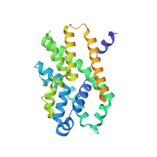
    Trimeric intracellular cation (TRIC) channels are thought to provide counter-ion currents that facilitate the active release of Ca 2+ from intracellular stores. TRIC activity is controlled by voltage and Ca 2+ modulation, but underlying mechanisms have remained unknown. Here we describe high-resolution crystal structures of vertebrate TRIC-A and TRIC-B channels, both in Ca 2+ -bound and Ca 2+ -free states, and we analyze conductance properties in structure-inspired mutagenesis experiments. The TRIC channels are symmetric trimers, wherein we find a pore in each protomer that is gated by a highly conserved lysine residue. In the resting state, Ca 2+ binding at the luminal surface of TRIC-A, on its threefold axis, stabilizes lysine blockage of the pores. During active Ca 2+ release, luminal Ca 2+ depletion removes inhibition to permit the lysine-bearing and voltage-sensing helix to move in response to consequent membrane hyperpolarization. Diacylglycerol is found at interprotomer interfaces, suggesting a role in metabolic control.


  • Organizational Affiliation
    • State Key Laboratory of Molecular Developmental Biology, Institute of Genetics and Developmental Biology, Chinese Academy of Sciences, Beijing 100101, China.

Macromolecule Content 

  • Total Structure Weight: 35.46 kDa 
  • Atom Count: 1,769 
  • Modeled Residue Count: 224 
  • Deposited Residue Count: 314 
  • Unique protein chains: 1

Macromolecules

Find similar proteins by:|  3D Structure
Entity ID: 1
MoleculeChains  Sequence LengthOrganismDetailsImage
Trimeric intracellular cation channel type B-B314Xenopus laevisMutation(s): 0 
Gene Names: tmem38b-b
Membrane Entity: Yes 
UniProt
Find proteins for Q6GN30 (Xenopus laevis)
Explore Q6GN30 
Go to UniProtKB:  Q6GN30
Entity Groups
Sequence Clusters30% Identity50% Identity70% Identity90% Identity95% Identity100% Identity
UniProt GroupQ6GN30
Sequence Annotations
Expand
Reference Sequence

Experimental Data & Validation

Experimental Data

  • Method: X-RAY DIFFRACTION
  • Resolution: 3.70 Å
  • R-Value Free:  0.327 (Depositor), 0.334 (DCC) 
  • R-Value Work:  0.311 (Depositor), 0.319 (DCC) 
  • R-Value Observed: 0.312 (Depositor) 
Space Group: F 4 3 2
Unit Cell:
Length ( Å )Angle ( ˚ )
a = 250.009α = 90
b = 250.009β = 90
c = 250.009γ = 90
Software Package:
Software NamePurpose
PHENIXrefinement
Aimlessdata scaling
PDB_EXTRACTdata extraction
XDSdata reduction
PHENIXphasing

Structure Validation

View Full Validation Report


Entry History 

& Funding Information

Deposition Data


Funding OrganizationLocationGrant Number
Ministry of Science and Technology (China)China2016YFA0500503
Ministry of Science and Technology (China)China2015CB910102

Revision History  (Full details and data files)

  • Version 1.0: 2019-05-01
    Type: Initial release
  • Version 1.1: 2019-11-06
    Changes: Data collection, Database references
  • Version 1.2: 2023-11-22
    Changes: Data collection, Database references, Refinement description