6IYA | pdb_00006iya

Structure of the DNA binding domain of antitoxin CopASO


Experimental Data Snapshot

  • Method: X-RAY DIFFRACTION
  • Resolution: 3.00 Å
  • R-Value Free: 
    0.298 (Depositor), 0.297 (DCC) 
  • R-Value Work: 
    0.247 (Depositor), 0.249 (DCC) 
  • R-Value Observed: 
    0.249 (Depositor) 

Starting Model: experimental
View more details

wwPDB Validation 3D Report Full Report

Validation slider image for 6IYA

This is version 1.2 of the entry. See complete history

Literature

Structure and allosteric coupling of type II antitoxin CopASO.

Zhao, R.Li, Q.Zhang, J.Li, F.Yao, J.Zhang, J.Liu, L.Wang, X.Zhang, X.

(2019) Biochem Biophys Res Commun 514: 1122-1127

  • DOI: https://doi.org/10.1016/j.bbrc.2019.05.049
  • Primary Citation Related Structures: 
    6IYA

  • PubMed Abstract: 

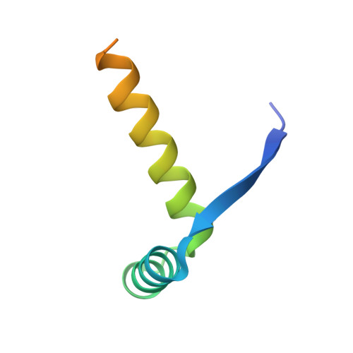
    Toxin-antitoxin (TA) systems play critical roles in the environment adaptation of bacteria. Allosteric coupling between the N-terminal DNA-binding domain and the C-terminal toxin-binding domain of antitoxins contributes to conditional cooperativity in the functioning of type II TA. Herein, using circular dichroism (CD), nuclear magnetic resonance (NMR), X-ray crystallography, and size exclusion chromatography (SEC), the structure and DNA binding of CopA SO , a newly identified type II antitoxin in Shewanella oneidensis, were investigated. Our data show that CopA SO is a typical RHH antitoxin with an ordered N-terminal domain and a disordered C-terminal domain, and furthermore indicate that the C-terminal domain facilitates DNA binding of the N-terminal domain, which in turn induces the C-terminal domain to fold and associate.


  • Organizational Affiliation
    • School of Life Sciences, Anhui University, Hefei, Anhui, 230601, China; Anhui Provincial Engineering Technology Research Center of Microorganisms and Biocatalysis, Hefei, Anhui, 230601, China; Anhui Key Laboratory of Modern Biomanufacturing, Hefei, Anhui, 230601, China.

Macromolecule Content 

  • Total Structure Weight: 47.96 kDa 
  • Atom Count: 2,478 
  • Modeled Residue Count: 286 
  • Deposited Residue Count: 396 
  • Unique protein chains: 1

Macromolecules

Find similar proteins by:|  3D Structure
Entity ID: 1
MoleculeChains  Sequence LengthOrganismDetailsImage
Transcriptional regulator CopG family
A, B, C, D, E
A, B, C, D, E, F
66Shewanella oneidensis MR-1Mutation(s): 0 
Gene Names: SO_1445
UniProt
Find proteins for Q8EGZ2 (Shewanella oneidensis (strain ATCC 700550 / JCM 31522 / CIP 106686 / LMG 19005 / NCIMB 14063 / MR-1))
Explore Q8EGZ2 
Go to UniProtKB:  Q8EGZ2
Entity Groups
Sequence Clusters30% Identity50% Identity70% Identity90% Identity95% Identity100% Identity
UniProt GroupQ8EGZ2
Sequence Annotations
Expand
Reference Sequence

Experimental Data & Validation

Experimental Data

  • Method: X-RAY DIFFRACTION
  • Resolution: 3.00 Å
  • R-Value Free:  0.298 (Depositor), 0.297 (DCC) 
  • R-Value Work:  0.247 (Depositor), 0.249 (DCC) 
  • R-Value Observed: 0.249 (Depositor) 
Space Group: P 21 2 21
Unit Cell:
Length ( Å )Angle ( ˚ )
a = 58.541α = 90
b = 60.949β = 90
c = 105.098γ = 90
Software Package:
Software NamePurpose
HKL-2000data reduction
HKL-2000data scaling
PHASERphasing
PHENIXrefinement
PDB_EXTRACTdata extraction

Structure Validation

View Full Validation Report


Entry History 

& Funding Information

Deposition Data


Funding OrganizationLocationGrant Number
National Natural Science Foundation of ChinaChina31470775

Revision History  (Full details and data files)

  • Version 1.0: 2019-01-16
    Type: Initial release
  • Version 1.1: 2019-08-07
    Changes: Data collection, Database references
  • Version 1.2: 2023-11-22
    Changes: Data collection, Database references, Refinement description