


Crystal structure of enoyl-CoA hydratase from Ralstonia eutropha H16
Son, H.F., Kim, K.J.To be published.
Experimental Data Snapshot
Starting Model: experimental
View more details
Entity ID: 1 | |||||
|---|---|---|---|---|---|
| Molecule | Chains | Sequence Length | Organism | Details | Image |
| Enoyl-CoA hydratase/Delta(3)-cis-delta(2)-trans-enoyl-CoA isomerase | A [auth B], B [auth A] | 701 | Cupriavidus necator H16 | Mutation(s): 0 Gene Names: H16_A1526 | 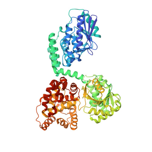 |
UniProt | |||||
Entity Groups | |||||
| Sequence Clusters | 30% Identity50% Identity70% Identity90% Identity95% Identity100% Identity | ||||
| UniProt Group | Q0KBG3 | ||||
Sequence AnnotationsExpand | |||||
Reference Sequence | |||||
| Ligands 2 Unique | |||||
|---|---|---|---|---|---|
| ID | Chains | Name / Formula / InChI Key | 2D Diagram | 3D Interactions | |
| NAD (Subject of Investigation/LOI) Download:Ideal Coordinates CCD File | F [auth B], J [auth A] | NICOTINAMIDE-ADENINE-DINUCLEOTIDE C21 H27 N7 O14 P2 BAWFJGJZGIEFAR-NNYOXOHSSA-N |  | ||
| GOL Download:Ideal Coordinates CCD File | C [auth B] D [auth B] E [auth B] G [auth A] H [auth A] | GLYCEROL C3 H8 O3 PEDCQBHIVMGVHV-UHFFFAOYSA-N |  | ||
| Length ( Å ) | Angle ( ˚ ) |
|---|---|
| a = 89.471 | α = 90 |
| b = 102.169 | β = 106.63 |
| c = 96.672 | γ = 90 |
| Software Name | Purpose |
|---|---|
| HKL-2000 | data reduction |
| REFMAC | refinement |
| PDB_EXTRACT | data extraction |
| HKL-2000 | data scaling |
| MOLREP | phasing |


