6IST | pdb_00006ist

Crystal structure of a wild type endolysin LysIME-EF1


Experimental Data Snapshot

  • Method: X-RAY DIFFRACTION
  • Resolution: 1.75 Å
  • R-Value Free: 
    0.223 (Depositor), 0.230 (DCC) 
  • R-Value Work: 
    0.180 (Depositor), 0.187 (DCC) 
  • R-Value Observed: 
    0.182 (Depositor) 

wwPDB Validation 3D Report Full Report

Validation slider image for 6IST

This is version 1.4 of the entry. See complete history

Literature

Structural and functional insights into a novel two-component endolysin encoded by a single gene in Enterococcus faecalis phage.

Zhou, B.Zhen, X.Zhou, H.Zhao, F.Fan, C.Perculija, V.Tong, Y.Mi, Z.Ouyang, S.

(2020) PLoS Pathog 16: e1008394-e1008394

  • DOI: https://doi.org/10.1371/journal.ppat.1008394
  • Primary Citation Related Structures: 
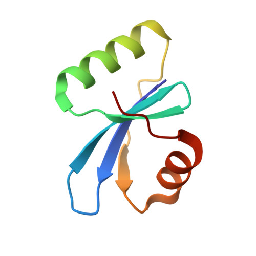
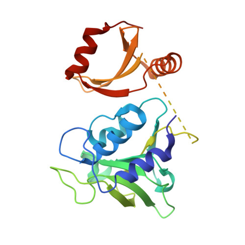
    6IST, 6L00

  • PubMed Abstract: 

    Using bacteriophage-derived endolysins as an alternative strategy for fighting drug-resistant bacteria has recently been garnering renewed interest. However, their application is still hindered by their narrow spectra of activity. In our previous work, we demonstrated that the endolysin LysIME-EF1 possesses efficient bactericidal activity against multiple strains of Enterococcus faecalis (E. faecalis). Herein, we observed an 8 kDa fragment and hypothesized that it contributes to LysIME-EF1 lytic activity. To examine our hypothesis, we determined the structure of LysIME-EF1 at 1.75 Å resolution. LysIME-EF1 exhibits a unique architecture in which one full-length LysIME-EF1 forms a tetramer with three additional C-terminal cell-wall binding domains (CBDs) that correspond to the abovementioned 8 kDa fragment. Furthermore, we identified an internal ribosomal binding site (RBS) and alternative start codon within LysIME-EF1 gene, which are demonstrated to be responsible for the translation of the truncated CBD. To elucidate the molecular mechanism for the lytic activity of LysIME-EF1, we combined mutagenesis, lytic activity assays and in vivo animal infection experiments. The results confirmed that the additional LysIME-EF1 CBDs are important for LysIME-EF1 architecture and its lytic activity. To our knowledge, this is the first determined structure of multimeric endolysin encoded by a single gene in E. faecalis phages. As such, it may provide valuable insights into designing potent endolysins against the opportunistic pathogen E. faecalis.


  • Organizational Affiliation
    • The Key Laboratory of Innate Immune Biology of Fujian Province, Provincial University Key Laboratory of Cellular Stress Response and Metabolic Regulation, Biomedical Research Center of South China, Key Laboratory of OptoElectronic Science and Technology for Medicine of the Ministry of Education, College of Life Sciences, Fujian Normal University, Fuzhou, China.

Macromolecule Content 

  • Total Structure Weight: 50.82 kDa 
  • Atom Count: 3,791 
  • Modeled Residue Count: 423 
  • Deposited Residue Count: 447 
  • Unique protein chains: 2

Macromolecules

Find similar proteins by:|  3D Structure
Entity ID: 1
MoleculeChains  Sequence LengthOrganismDetailsImage
Lysin
A, B, D
70Enterococcus phage IMEEF1Mutation(s): 0 
UniProt
Find proteins for S5MRN1 (Enterococcus phage IMEEF1)
Explore S5MRN1 
Go to UniProtKB:  S5MRN1
Entity Groups
Sequence Clusters30% Identity50% Identity70% Identity90% Identity95% Identity100% Identity
UniProt GroupS5MRN1
Sequence Annotations
Expand
Reference Sequence
Find similar proteins by:|  3D Structure
Entity ID: 2
MoleculeChains  Sequence LengthOrganismDetailsImage
Lysin237Enterococcus phage IMEEF1Mutation(s): 0 
UniProt
Find proteins for S5MRN1 (Enterococcus phage IMEEF1)
Explore S5MRN1 
Go to UniProtKB:  S5MRN1
Entity Groups
Sequence Clusters30% Identity50% Identity70% Identity90% Identity95% Identity100% Identity
UniProt GroupS5MRN1
Sequence Annotations
Expand
Reference Sequence

Small Molecules

Ligands 1 Unique
IDChains Name / Formula / InChI Key2D Diagram3D Interactions
CA

Query on CA


Download:Ideal Coordinates CCD File
E [auth C]CALCIUM ION
Ca
BHPQYMZQTOCNFJ-UHFFFAOYSA-N

Experimental Data & Validation

Experimental Data

  • Method: X-RAY DIFFRACTION
  • Resolution: 1.75 Å
  • R-Value Free:  0.223 (Depositor), 0.230 (DCC) 
  • R-Value Work:  0.180 (Depositor), 0.187 (DCC) 
  • R-Value Observed: 0.182 (Depositor) 
Space Group: P 1 21 1
Unit Cell:
Length ( Å )Angle ( ˚ )
a = 47.03α = 90
b = 56.92β = 96.29
c = 91.52γ = 90
Software Package:
Software NamePurpose
REFMACrefinement
HKL-2000data reduction
HKL-2000data scaling
PHASERphasing

Structure Validation

View Full Validation Report


Entry History 

Deposition Data

  • Released Date: 2019-11-20 
  • Deposition Author(s): Ouyang, S.Y.

Revision History  (Full details and data files)

  • Version 1.0: 2019-11-20
    Type: Initial release
  • Version 1.1: 2020-02-12
    Changes: Experimental preparation
  • Version 1.2: 2020-02-19
    Changes: Database references, Structure summary
  • Version 1.3: 2020-04-15
    Changes: Database references
  • Version 1.4: 2024-03-27
    Changes: Data collection, Database references