6IRE | pdb_00006ire

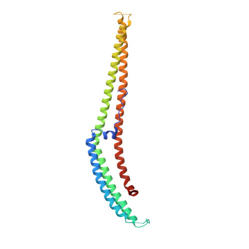
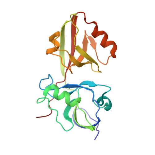
Complex structure of INAD PDZ45 and NORPA CC-PBM


Experimental Data Snapshot

  • Method: X-RAY DIFFRACTION
  • Resolution: 3.25 Å
  • R-Value Free: 
    0.275 (Depositor), 0.273 (DCC) 
  • R-Value Work: 
    0.225 (Depositor), 0.227 (DCC) 
  • R-Value Observed: 
    0.228 (Depositor) 

wwPDB Validation 3D Report Full Report

Validation slider image for 6IRE

This is version 1.2 of the entry. See complete history. 

Literature

An unexpected INAD PDZ tandem-mediated plc beta binding in Drosophila photo receptors.

Ye, F., Huang, Y., Li, J., Ma, Y., Xie, C., Liu, Z., Deng, X., Wan, J., Xue, T., Liu, W., Zhang, M.

(2018) Elife 7

  • DOI: https://doi.org/10.7554/eLife.41848
  • Primary Citation Related Structures: 
    6IRB, 6IRC, 6IRD, 6IRE

  • PubMed Abstract: 

    INAD assembles key enzymes of the Drosophila compound eye photo-transduction pathway into a supramolecular complex, supporting efficient and fast light signaling. However, the molecular mechanism that governs the interaction between INAD and NORPA (phospholipase Cβ, PLCβ), a key step for the fast kinetics of the light signaling, is not known. Here, we show that the NORPA C-terminal coiled-coil domain and PDZ-binding motif (CC-PBM) synergistically bind to INAD PDZ45 tandem with an unexpected mode and unprecedented high affinity. Guided by the structure of the INAD-NORPA complex, we discover that INADL is probably a mammalian counterpart of INAD. The INADL PDZ89 tandem specifically binds to PLCβ4 with a mode that is strikingly similar to that of the INAD-NORPA complex, as revealed by the structure of the INADL PDZ89-PLCβ4 CC-PBM complex. Therefore, our study suggests that the highly specific PDZ tandem - PLCβ interactions are an evolutionarily conserved mechanism in PLCβ signaling in the animal kingdom.


  • Organizational Affiliation: 
    • Division of Life Science, State Key Laboratory of Molecular Neuroscience, Hong Kong University of Science and Technology, Hong Kong, China.

Macromolecule Content 

  • Total Structure Weight: 48.39 kDa 
  • Atom Count: 2,982 
  • Modeled Residue Count: 416 
  • Deposited Residue Count: 428 
  • Unique protein chains: 2

Macromolecules

Find similar proteins by:|  3D Structure
Entity ID: 1
MoleculeChains  Sequence LengthOrganismDetailsImage
1-phosphatidylinositol 4,5-bisphosphate phosphodiesterase234Drosophila melanogasterMutation(s): 8 
Gene Names: norpA, PLC-beta, CG3620
EC: 3.1.4.11
UniProt
Find proteins for P13217 (Drosophila melanogaster)
Explore P13217 
Go to UniProtKB:  P13217
Entity Groups
Sequence Clusters30% Identity50% Identity70% Identity90% Identity95% Identity100% Identity
UniProt GroupP13217
Sequence Annotations
Expand
Reference Sequence
Find similar proteins by:|  3D Structure
Entity ID: 2
MoleculeChains  Sequence LengthOrganismDetailsImage
Inactivation-no-after-potential D protein194Drosophila melanogasterMutation(s): 0 
Gene Names: inaD, CG3504
UniProt
Find proteins for Q24008 (Drosophila melanogaster)
Explore Q24008 
Go to UniProtKB:  Q24008
Entity Groups
Sequence Clusters30% Identity50% Identity70% Identity90% Identity95% Identity100% Identity
UniProt GroupQ24008
Sequence Annotations
Expand
Reference Sequence

Experimental Data & Validation

Experimental Data

  • Method: X-RAY DIFFRACTION
  • Resolution: 3.25 Å
  • R-Value Free:  0.275 (Depositor), 0.273 (DCC) 
  • R-Value Work:  0.225 (Depositor), 0.227 (DCC) 
  • R-Value Observed: 0.228 (Depositor) 
Space Group: C 2 2 21
Unit Cell:
Length ( Å )Angle ( ˚ )
a = 117.946α = 90
b = 157.213β = 90
c = 60.986γ = 90
Software Package:
Software NamePurpose
PHENIXrefinement
DENZOdata reduction
SCALEPACKdata scaling
PDB_EXTRACTdata extraction
PHASERphasing

Structure Validation

View Full Validation Report


Entry History 

Deposition Data

Revision History  (Full details and data files)

  • Version 1.0: 2019-01-23
    Type: Initial release
  • Version 1.1: 2020-10-28
    Changes: Database references
  • Version 1.2: 2024-03-27
    Changes: Data collection, Database references