6IQZ

High resolution structure of bilirubin oxidase from Myrothecium verrucaria - wild type


Experimental Data Snapshot

  • Method: X-RAY DIFFRACTION
  • Resolution: 1.46 Å
  • R-Value Free: 0.170 
  • R-Value Work: 0.131 
  • R-Value Observed: 0.132 

wwPDB Validation   3D Report Full Report


Ligand Structure Quality Assessment 


This is version 2.1 of the entry. See complete history


Literature

Redox Potential-Dependent Formation of an Unusual His-Trp Bond in Bilirubin Oxidase.

Akter, M.Tokiwa, T.Shoji, M.Nishikawa, K.Shigeta, Y.Sakurai, T.Higuchi, Y.Kataoka, K.Shibata, N.

(2018) Chemistry 24: 18052-18058

  • DOI: https://doi.org/10.1002/chem.201803798
  • Primary Citation of Related Structures:  
    6IQX, 6IQY, 6IQZ

  • PubMed Abstract: 

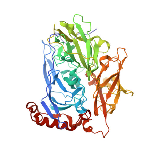
    Bilirubin oxidase (BOD) belongs to the family of blue multicopper oxidases, and catalyzes the concomitant oxidation of bilirubin to biliverdin and the reduction of molecular oxygen to water via a four-electron reduction system. The active sites of BOD comprise four copper atoms; type I copper (T1Cu) forms a mononuclear site, and a cluster of three copper atoms forms a trinuclear center. In the present study, we determined the high-resolution crystal structures of BOD from the fungus Myrothecium verrucaria. We investigated wild-type (WT) BOD and a BOD mutant called Met467Gln, which is inactive against bilirubin. The structures revealed that a novel post-translational crosslink between Trp396 and His398 is formed in the vicinity of the T1Cu site in WT BOD, whereas it is absent in the Met467Gln mutant. Our structural and computational studies suggest that the His-Trp crosslink adjusts the redox potential of T1Cu to that of bilirubin to efficiently abstract electrons from the substrate.


  • Organizational Affiliation

    Department of Picobiology, Graduate School of Life Science, University of Hyogo, 3-2-1 Koto, Kamigori-cho, Ako-gun, Hyogo, 678-1297, Japan.


Macromolecules
Find similar proteins by:  (by identity cutoff)  |  3D Structure
Entity ID: 1
MoleculeChains Sequence LengthOrganismDetailsImage
Bilirubin oxidase538Albifimbria verrucariaMutation(s): 0 
EC: 1.3.3.5
UniProt
Find proteins for Q12737 (Albifimbria verrucaria)
Explore Q12737 
Go to UniProtKB:  Q12737
Entity Groups  
Sequence Clusters30% Identity50% Identity70% Identity90% Identity95% Identity100% Identity
UniProt GroupQ12737
Sequence Annotations
Expand
  • Reference Sequence
Oligosaccharides

Help

Entity ID: 2
MoleculeChains Length2D Diagram Glycosylation3D Interactions
alpha-D-mannopyranose-(1-4)-2-acetamido-2-deoxy-beta-D-glucopyranose-(1-4)-2-acetamido-2-deoxy-beta-D-glucopyranose
B
3N-Glycosylation
Glycosylation Resources
GlyTouCan:  G62182OO
GlyCosmos:  G62182OO
GlyGen:  G62182OO
Experimental Data & Validation

Experimental Data

  • Method: X-RAY DIFFRACTION
  • Resolution: 1.46 Å
  • R-Value Free: 0.170 
  • R-Value Work: 0.131 
  • R-Value Observed: 0.132 
  • Space Group: C 1 2 1
Unit Cell:
Length ( Å )Angle ( ˚ )
a = 138.342α = 90
b = 83.553β = 95.73
c = 52.69γ = 90
Software Package:
Software NamePurpose
HKL-2000data reduction
HKL-2000data scaling
PHENIXphasing
PHENIXrefinement

Structure Validation

View Full Validation Report


Ligand Structure Quality Assessment 


Entry History 

Deposition Data

Revision History  (Full details and data files)

  • Version 1.0: 2018-11-21
    Type: Initial release
  • Version 1.1: 2018-12-26
    Changes: Data collection, Database references
  • Version 2.0: 2020-07-29
    Type: Remediation
    Reason: Carbohydrate remediation
    Changes: Atomic model, Data collection, Derived calculations, Structure summary
  • Version 2.1: 2023-11-22
    Changes: Data collection, Database references, Refinement description, Structure summary