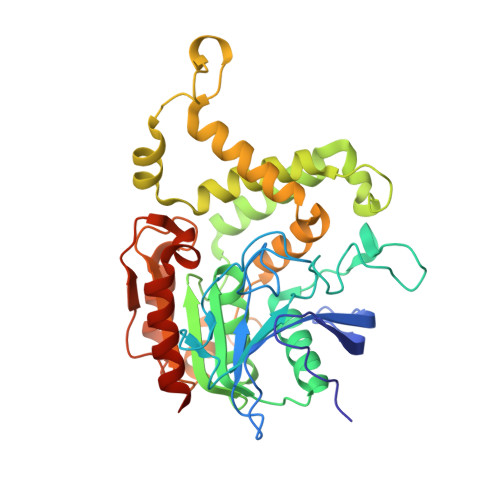
6IOG

Crystal structure of Homoserine O-acetyltransferase from Mycobacterium smegmatis ATCC 19420


Experimental Data Snapshot

  • Method: X-RAY DIFFRACTION
  • Resolution: 1.55 Å
  • R-Value Free: 0.196 
  • R-Value Work: 0.172 
  • R-Value Observed: 0.173 

Starting Model: experimental
View more details

wwPDB Validation   3D Report Full Report


This is version 1.1 of the entry. See complete history


Literature

Crystal structure and biochemical characterization of O-acetylhomoserine acetyltransferase from Mycobacterium smegmatis ATCC 19420.

Sagong, H.Y.Hong, J.Kim, K.J.

(2019) Biochem Biophys Res Commun 517: 399-406

  • DOI: https://doi.org/10.1016/j.bbrc.2019.07.117
  • Primary Citation of Related Structures:  
    6IOG, 6IOH, 6IOI

  • PubMed Abstract: 

    Mycobacterium smegmatis is a good model for studying the physiology and pathogenesis of Mycobacterium tuberculosis due to its genetic similarity. As methionine biosynthesis exists only in microorganisms, the enzymes involved in methionine biosynthesis can be a potential target for novel antibiotics. Homoserine O-acetyltransferase from M. smegmatis (MsHAT) catalyzes the transfer of acetyl-group from acetyl-CoA to homoserine. To investigate the molecular mechanism of MsHAT, we determined its crystal structure in apo-form and in complex with either CoA or homoserine and revealed the substrate binding mode of MsHAT. A structural comparison of MsHAT with other HATs suggests that the conformation of the α5 to α6 region might influence the shape of the dimer. In addition, the active site entrance shows an open or closed conformation and might determine the substrate binding affinity of HATs.


  • Organizational Affiliation

    School of Life Sciences (KNU Creative BioResearch Group), KNU Institute for Microorganisms, Kyungpook National University, Daegu, 41566, Republic of Korea.


Macromolecules
Find similar proteins by:  (by identity cutoff)  |  3D Structure
Entity ID: 1
MoleculeChains Sequence LengthOrganismDetailsImage
Homoserine O-acetyltransferase
A, B
374Mycolicibacterium smegmatisMutation(s): 0 
Gene Names: metX_1metXAERS451418_01697
EC: 2.3.1.31
UniProt
Find proteins for A0QSZ0 (Mycolicibacterium smegmatis (strain ATCC 700084 / mc(2)155))
Explore A0QSZ0 
Go to UniProtKB:  A0QSZ0
Entity Groups  
Sequence Clusters30% Identity50% Identity70% Identity90% Identity95% Identity100% Identity
UniProt GroupA0QSZ0
Sequence Annotations
Expand
  • Reference Sequence
Small Molecules
Ligands 1 Unique
IDChains Name / Formula / InChI Key2D Diagram3D Interactions
GOL
Query on GOL

Download Ideal Coordinates CCD File 
C [auth A]GLYCEROL
C3 H8 O3
PEDCQBHIVMGVHV-UHFFFAOYSA-N
Experimental Data & Validation

Experimental Data

  • Method: X-RAY DIFFRACTION
  • Resolution: 1.55 Å
  • R-Value Free: 0.196 
  • R-Value Work: 0.172 
  • R-Value Observed: 0.173 
  • Space Group: P 21 21 21
Unit Cell:
Length ( Å )Angle ( ˚ )
a = 58.357α = 90
b = 96.246β = 90
c = 140.351γ = 90
Software Package:
Software NamePurpose
HKL-2000data reduction
REFMACrefinement
PDB_EXTRACTdata extraction
HKL-2000data scaling
MOLREPphasing

Structure Validation

View Full Validation Report


Entry History 

Deposition Data

Revision History  (Full details and data files)

  • Version 1.0: 2019-09-04
    Type: Initial release
  • Version 1.1: 2023-11-22
    Changes: Data collection, Database references, Refinement description