6INZ | pdb_00006inz

Crystal structure of solute-binding protein complexed with unsaturated hyaluronan disaccharide


Experimental Data Snapshot

  • Method: X-RAY DIFFRACTION
  • Resolution: 2.29 Å
  • R-Value Free: 
    0.217 (Depositor), 0.219 (DCC) 
  • R-Value Work: 
    0.160 (Depositor), 0.162 (DCC) 
  • R-Value Observed: 
    0.162 (Depositor) 

Starting Model: experimental
View more details

wwPDB Validation 3D Report Full Report

Validation slider image for 6INZ

This is version 2.1 of the entry. See complete history

Literature

Substrate recognition by bacterial solute-binding protein is responsible for import of extracellular hyaluronan and chondroitin sulfate from the animal host.

Oiki, S.Sato, M.Mikami, B.Murata, K.Hashimoto, W.

(2019) Biosci Biotechnol Biochem 83: 1946-1954

  • DOI: https://doi.org/10.1080/09168451.2019.1630250
  • Primary Citation Related Structures: 
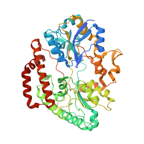
    6INZ

  • PubMed Abstract: 

    Glycosaminoglycans (GAGs) such as hyaluronan and chondroitin in animal extracellular matrices contain disaccharide-repeating units. In a Gram-negative pathogenic Streptobacillus moniliformis , which belongs to Fusobacteria phylum and resides in rodent oral cavities, the solute-binding protein (Smon0123)-dependent ATP-binding cassette transporter imports unsaturated hyaluronan/chondroitin disaccharides into the cytoplasm after GAG lyase-dependent depolymerization. Here we show substrate recognition of unsaturated hyaluronan disaccharide by Smon0123. Moreover, Smon0123 exhibited no affinity for unsaturated chondroitin disaccharides containing three sulfate groups, distinct from non-sulfated, mono-sulfated, and di-sulfated chondroitin disaccharides previously identified as substrates. Crystal structure of Smon0123 with unsaturated hyaluronan disaccharide demonstrates that several residues, including Trp284 and Glu410, are crucial for binding to unsaturated hyaluronan/chondroitin disaccharides, whereas arrangements of water molecules at binding sites are found to be substrate dependent through comparison with substrate-bound structures determined previously. These residues are well conserved in Smon0123-like proteins of fusobacteria, and probably facilitate the fusobacterial residence in hyaluronan-rich oral cavities.


  • Organizational Affiliation
    • Laboratory of Basic and Applied Molecular Biotechnology, Division of Food Science and Biotechnology, Graduate School of Agriculture, Kyoto University , Kyoto , Japan.

Macromolecule Content 

  • Total Structure Weight: 58.72 kDa 
  • Atom Count: 4,232 
  • Modeled Residue Count: 474 
  • Deposited Residue Count: 505 
  • Unique protein chains: 1

Macromolecules

Find similar proteins by:|  3D Structure
Entity ID: 1
MoleculeChains  Sequence LengthOrganismDetailsImage
Extracellular solute-binding protein family 1505Streptobacillus moniliformis DSM 12112Mutation(s): 0 
Gene Names: Smon_0123
UniProt
Find proteins for D1AWE0 (Streptobacillus moniliformis (strain ATCC 14647 / DSM 12112 / NCTC 10651 / 9901))
Explore D1AWE0 
Go to UniProtKB:  D1AWE0
Entity Groups
Sequence Clusters30% Identity50% Identity70% Identity90% Identity95% Identity100% Identity
UniProt GroupD1AWE0
Sequence Annotations
Expand
Reference Sequence

Oligosaccharides

Help  
Entity ID: 2
MoleculeChains Length2D Diagram GlycosylationD Interactions
4-deoxy-alpha-L-threo-hex-4-enopyranuronic acid-(1-3)-2-acetamido-2-deoxy-beta-D-glucopyranose
B
2N/A
Glycosylation Resources
GlyTouCan: G14110BE
GlyCosmos: G14110BE

Small Molecules

Ligands 2 Unique
IDChains Name / Formula / InChI Key2D Diagram3D Interactions
SO4

Query on SO4


Download:Ideal Coordinates CCD File
C [auth A]
D [auth A]
E [auth A]
F [auth A]
G [auth A]
C [auth A],
D [auth A],
E [auth A],
F [auth A],
G [auth A],
H [auth A],
I [auth A],
J [auth A]
SULFATE ION
O4 S
QAOWNCQODCNURD-UHFFFAOYSA-L
CA

Query on CA


Download:Ideal Coordinates CCD File
K [auth A]CALCIUM ION
Ca
BHPQYMZQTOCNFJ-UHFFFAOYSA-N

Experimental Data & Validation

Experimental Data

  • Method: X-RAY DIFFRACTION
  • Resolution: 2.29 Å
  • R-Value Free:  0.217 (Depositor), 0.219 (DCC) 
  • R-Value Work:  0.160 (Depositor), 0.162 (DCC) 
  • R-Value Observed: 0.162 (Depositor) 
Space Group: P 21 21 21
Unit Cell:
Length ( Å )Angle ( ˚ )
a = 48.74α = 90
b = 97.501β = 90
c = 132.443γ = 90
Software Package:
Software NamePurpose
PHENIXrefinement
HKL-2000data collection
MOLREPphasing
DENZOdata reduction
SCALEPACKdata scaling

Structure Validation

View Full Validation Report


Entry History 

& Funding Information

Deposition Data


Funding OrganizationLocationGrant Number
Japan Society for the Promotion of ScienceJapan--

Revision History  (Full details and data files)

  • Version 1.0: 2019-09-11
    Type: Initial release
  • Version 2.0: 2020-07-29
    Type: Remediation
    Reason: Carbohydrate remediation
    Changes: Atomic model, Data collection, Derived calculations, Structure summary
  • Version 2.1: 2023-11-22
    Changes: Data collection, Database references, Derived calculations, Refinement description, Structure summary