6IFA

Structure of beta-trefoil lectin from Entamoeba histolytica


Experimental Data Snapshot

  • Method: X-RAY DIFFRACTION
  • Resolution: 1.90 Å
  • R-Value Free: 0.236 
  • R-Value Work: 0.194 
  • R-Value Observed: 0.196 

wwPDB Validation   3D Report Full Report


This is version 1.1 of the entry. See complete history


Literature

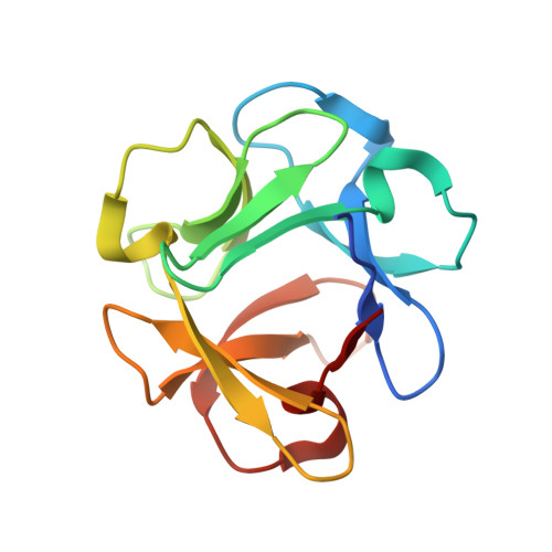
Crystal structures of a beta-trefoil lectin from Entamoeba histolytica in monomeric and a novel disulphide bond-mediated dimeric forms.

Khan, F.Kurre, D.Suguna, K.

(2020) Glycobiology 

  • DOI: https://doi.org/10.1093/glycob/cwaa001
  • Primary Citation of Related Structures:  
    6IFA, 6IFB

  • PubMed Abstract: 

    β-Trefoil lectins are galactose/N-acetyl galactosamine specific lectins, which are widely distributed across all kingdoms of life and are known to perform several important functions. However, there is no report available on the characterization of these lectins from protozoans. We have performed structural and biophysical studies on a β-trefoil lectin from Entamoeba histolytica (EntTref), which exists as a mixture of monomers and dimers in solution. Further, we have determined the affinities of EntTref for rhamnose, galactose and different galactose-linked sugars. We obtained the crystal structure of EntTref in a sugar-free form (EntTref_apo) and a rhamnose-bound form (EntTref_rham). A novel Cys residue-mediated dimerization was revealed in the crystal structure of EntTref_apo while the structure of EntTref_rham provided the structural basis for the recognition of rhamnose by a β-trefoil lectin for the first time. To the best of our knowledge, this is the only report of the structural, functional and biophysical characterization of a β-trefoil lectin from a protozoan source and the first report of Cys-mediated dimerization in this class of lectins.


  • Organizational Affiliation

    Molecular Biophysics Unit, Indian Institute of Science, Bangalore, CV Raman Rd, 560012, India.


Macromolecules
Find similar proteins by:  (by identity cutoff)  |  3D Structure
Entity ID: 1
MoleculeChains Sequence LengthOrganismDetailsImage
lectin
A, B, C
129Entamoeba histolytica HM-1:IMSSMutation(s): 0 
UniProt
Find proteins for N9TFI9 (Entamoeba histolytica HM-1:IMSS-A)
Explore N9TFI9 
Go to UniProtKB:  N9TFI9
Entity Groups  
Sequence Clusters30% Identity50% Identity70% Identity90% Identity95% Identity100% Identity
UniProt GroupN9TFI9
Sequence Annotations
Expand
  • Reference Sequence
Small Molecules
Ligands 2 Unique
IDChains Name / Formula / InChI Key2D Diagram3D Interactions
GOL
Query on GOL

Download Ideal Coordinates CCD File 
D [auth A],
E [auth A],
H [auth A],
J [auth B],
O [auth C]
GLYCEROL
C3 H8 O3
PEDCQBHIVMGVHV-UHFFFAOYSA-N
IPA
Query on IPA

Download Ideal Coordinates CCD File 
F [auth A]
G [auth A]
I [auth A]
K [auth B]
L [auth B]
F [auth A],
G [auth A],
I [auth A],
K [auth B],
L [auth B],
M [auth B],
N [auth C],
P [auth C],
Q [auth C]
ISOPROPYL ALCOHOL
C3 H8 O
KFZMGEQAYNKOFK-UHFFFAOYSA-N
Experimental Data & Validation

Experimental Data

  • Method: X-RAY DIFFRACTION
  • Resolution: 1.90 Å
  • R-Value Free: 0.236 
  • R-Value Work: 0.194 
  • R-Value Observed: 0.196 
  • Space Group: I 4 2 2
Unit Cell:
Length ( Å )Angle ( ˚ )
a = 101.732α = 90
b = 101.732β = 90
c = 183.996γ = 90
Software Package:
Software NamePurpose
PHENIXrefinement
REFMACrefinement
XDSdata reduction
XDSdata scaling
PHASERphasing

Structure Validation

View Full Validation Report


Entry History 

Deposition Data

Revision History  (Full details and data files)

  • Version 1.0: 2019-09-25
    Type: Initial release
  • Version 1.1: 2020-04-08
    Changes: Database references