6IDP | pdb_00006idp

Crystal structure of Vibrio cholerae MATE transporter VcmN in the straight form


Experimental Data Snapshot

  • Method: X-RAY DIFFRACTION
  • Resolution: 2.21 Å
  • R-Value Free: 
    0.240 (Depositor), 0.243 (DCC) 
  • R-Value Work: 
    0.198 (Depositor), 0.201 (DCC) 
  • R-Value Observed: 
    0.201 (Depositor) 

wwPDB Validation 3D Report Full Report

Validation slider image for 6IDP

Ligand Structure Quality Assessment 


This is version 1.2 of the entry. See complete history

Literature

Structural Basis of H+-Dependent Conformational Change in a Bacterial MATE Transporter.

Kusakizako, T.Claxton, D.P.Tanaka, Y.Maturana, A.D.Kuroda, T.Ishitani, R.Mchaourab, H.S.Nureki, O.

(2019) Structure 27: 293

  • DOI: https://doi.org/10.1016/j.str.2018.10.004
  • Primary Citation Related Structures: 
    6IDP, 6IDR, 6IDS

  • PubMed Abstract: 

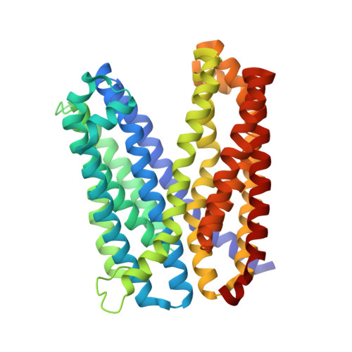
    Multidrug and toxic compound extrusion (MATE) transporters efflux toxic compounds using a Na + or H + gradient across the membrane. Although the structures of MATE transporters have been reported, the cation-coupled substrate transport mechanism remains controversial. Here we report crystal structures of VcmN, a Vibrio cholerae MATE transporter driven by the H + gradient. High-resolution structures in two distinct conformations associated with different pHs revealed that the rearrangement of the hydrogen-bonding network around the conserved Asp35 induces the bending of transmembrane helix 1, as in the case of the H + -coupled Pyrococcus furiosus MATE transporter. We also determined the crystal structure of the D35N mutant, which captured a unique conformation of TM1 facilitated by an altered hydrogen-bonding network. Based on the present results, we propose a common step in the transport cycle shared among prokaryotic H + -coupled MATE transporters.


  • Organizational Affiliation
    • Department of Biological Sciences, Graduate School of Science, The University of Tokyo, 7-3-1 Hongo, Bunkyo-ku, Tokyo 113-0033, Japan.

Macromolecule Content 

  • Total Structure Weight: 54.3 kDa 
  • Atom Count: 3,600 
  • Modeled Residue Count: 438 
  • Deposited Residue Count: 441 
  • Unique protein chains: 1

Macromolecules

Find similar proteins by:|  3D Structure
Entity ID: 1
MoleculeChains  Sequence LengthOrganismDetailsImage
MATE family efflux transporter441Vibrio choleraeMutation(s): 0 
Gene Names: CGT79_05080

Small Molecules

Ligands 1 Unique
IDChains Name / Formula / InChI Key2D Diagram3D Interactions
OLC

Query on OLC


Download:Ideal Coordinates CCD File
B [auth A]
C [auth A]
D [auth A]
E [auth A]
F [auth A]
B [auth A],
C [auth A],
D [auth A],
E [auth A],
F [auth A],
G [auth A],
H [auth A],
I [auth A],
J [auth A],
K [auth A],
L [auth A],
M [auth A],
N [auth A],
O [auth A],
P [auth A],
Q [auth A],
R [auth A],
S [auth A],
T [auth A]
(2R)-2,3-dihydroxypropyl (9Z)-octadec-9-enoate
C21 H40 O4
RZRNAYUHWVFMIP-GDCKJWNLSA-N

Experimental Data & Validation

Experimental Data

  • Method: X-RAY DIFFRACTION
  • Resolution: 2.21 Å
  • R-Value Free:  0.240 (Depositor), 0.243 (DCC) 
  • R-Value Work:  0.198 (Depositor), 0.201 (DCC) 
  • R-Value Observed: 0.201 (Depositor) 
Space Group: P 21 21 21
Unit Cell:
Length ( Å )Angle ( ˚ )
a = 62.225α = 90
b = 92.027β = 90
c = 101.302γ = 90
Software Package:
Software NamePurpose
XDSdata scaling
PHENIXrefinement
PDB_EXTRACTdata extraction
XDSdata reduction
PHASERphasing

Structure Validation

View Full Validation Report


Ligand Structure Quality Assessment 


Entry History 

Deposition Data

Revision History  (Full details and data files)

  • Version 1.0: 2019-01-16
    Type: Initial release
  • Version 1.1: 2019-02-20
    Changes: Data collection, Database references
  • Version 1.2: 2024-03-27
    Changes: Data collection, Database references