6IAF

Fifteen minutes iron loaded Rana Catesbeiana H' ferritin variant H54N


Experimental Data Snapshot

  • Method: X-RAY DIFFRACTION
  • Resolution: 1.35 Å
  • R-Value Free: 0.141 
  • R-Value Work: 0.113 
  • R-Value Observed: 0.115 

wwPDB Validation   3D Report Full Report


This is version 1.1 of the entry. See complete history


Literature

Effect of the point mutation H54N on the ferroxidase process of Rana catesbeiana H' ferritin.

Pozzi, C.Di Pisa, F.Lalli, D.Rosa, C.Turano, P.Mangani, S.

(2019) J Inorg Biochem 197: 110697-110697

  • DOI: https://doi.org/10.1016/j.jinorgbio.2019.110697
  • Primary Citation of Related Structures:  
    6I9P, 6I9T, 6IAF, 6IAJ

  • PubMed Abstract: 

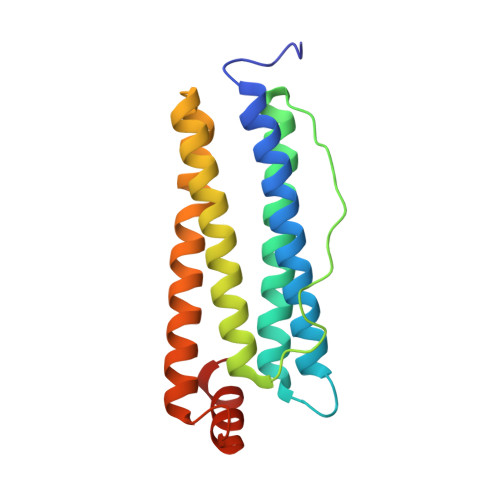
    Human H and Rana catesbeiana H' subunits in vertebrate ferritin protein cages catalyze the Fe(II) oxidation by molecular oxygen and promote the ferric oxide biomineral synthesis. By depositing iron biomineral, ferritins also prevent potentially toxic reactions products from Fe(II)-based Fenton chemistry. Recent work from our laboratory was aimed to describe the iron pathways within ferritin, from entrance into the cage to the ferroxidase site, and to understand the role played by amino-acid residues in iron trafficking and catalysis. Our approach exploits anomalous X-ray diffraction from ferritin crystals, exposed to a ferrous salt, to track transient iron binding sites along the path towards a well-defined di-iron site where they get oxidized by oxygen. Coupling structure determination with solution kinetic measurements on selected variants, allows validating the role played by key residues on the catalytic iron oxidation. Our previous studies on H' ferritin indicated the regulatory role played by His54, and by its human counterpart Gln58, on guiding Fe(II) ions to the catalytic site. Here, we have investigated the effects induced by substituting the wild type His54 with Asn54, having different iron coordination properties. We have obtained a series of atomic-resolution crystal structures that provide time-dependent snapshots of iron bound at different locations in the H' ferritin H54N variant. The comparison with H' ferritin and H' ferritin H54Q variant leads to identify a new iron binding site. Our kinetic and structural data support the role of H' ferritin residue 54 in regulating the access of Fe(II) ions to the catalytic site.


  • Organizational Affiliation

    Department of Biotechnology, Chemistry and Pharmacy, Department of Excellence 2018-2020, University of Siena, via Aldo Moro, 2, 53110 Siena, Italy. Electronic address: pozzi4@unisi.it.


Macromolecules
Find similar proteins by:  (by identity cutoff)  |  3D Structure
Entity ID: 1
MoleculeChains Sequence LengthOrganismDetailsImage
Ferritin, middle subunit176Aquarana catesbeianaMutation(s): 1 
EC: 1.16.3.1
UniProt
Find proteins for P07798 (Aquarana catesbeiana)
Explore P07798 
Go to UniProtKB:  P07798
Entity Groups  
Sequence Clusters30% Identity50% Identity70% Identity90% Identity95% Identity100% Identity
UniProt GroupP07798
Sequence Annotations
Expand
  • Reference Sequence
Small Molecules
Ligands 3 Unique
IDChains Name / Formula / InChI Key2D Diagram3D Interactions
FE2
Query on FE2

Download Ideal Coordinates CCD File 
B [auth A]
C [auth A]
D [auth A]
E [auth A]
F [auth A]
B [auth A],
C [auth A],
D [auth A],
E [auth A],
F [auth A],
G [auth A]
FE (II) ION
Fe
CWYNVVGOOAEACU-UHFFFAOYSA-N
CL
Query on CL

Download Ideal Coordinates CCD File 
O [auth A]
P [auth A]
Q [auth A]
R [auth A]
S [auth A]
O [auth A],
P [auth A],
Q [auth A],
R [auth A],
S [auth A],
T [auth A],
U [auth A],
V [auth A],
W [auth A]
CHLORIDE ION
Cl
VEXZGXHMUGYJMC-UHFFFAOYSA-M
MG
Query on MG

Download Ideal Coordinates CCD File 
H [auth A]
I [auth A]
J [auth A]
K [auth A]
L [auth A]
H [auth A],
I [auth A],
J [auth A],
K [auth A],
L [auth A],
M [auth A],
N [auth A]
MAGNESIUM ION
Mg
JLVVSXFLKOJNIY-UHFFFAOYSA-N
Experimental Data & Validation

Experimental Data

  • Method: X-RAY DIFFRACTION
  • Resolution: 1.35 Å
  • R-Value Free: 0.141 
  • R-Value Work: 0.113 
  • R-Value Observed: 0.115 
  • Space Group: F 4 3 2
Unit Cell:
Length ( Å )Angle ( ˚ )
a = 183.72α = 90
b = 183.72β = 90
c = 183.72γ = 90
Software Package:
Software NamePurpose
REFMACrefinement
MOSFLMdata reduction
XDSdata reduction
SCALAdata scaling
MOLREPphasing

Structure Validation

View Full Validation Report


Entry History 

Deposition Data

Revision History  (Full details and data files)

  • Version 1.0: 2019-05-22
    Type: Initial release
  • Version 1.1: 2024-01-24
    Changes: Data collection, Database references, Derived calculations, Refinement description