6IA6 | pdb_00006ia6

Crystal structure of the bacterial Dehalococcoides mccartyi Elp3 with desulfo-CoA


Experimental Data Snapshot

  • Method: X-RAY DIFFRACTION
  • Resolution: 2.70 Å
  • R-Value Free: 
    0.269 (Depositor), 0.272 (DCC) 
  • R-Value Work: 
    0.223 (Depositor), 0.231 (DCC) 
  • R-Value Observed: 
    0.225 (Depositor) 

Starting Model: experimental
View more details

wwPDB Validation 3D Report Full Report

Validation slider image for 6IA6

Ligand Structure Quality Assessment 


This is version 1.1 of the entry. See complete history

Literature

The Elongator subunit Elp3 is a non-canonical tRNA acetyltransferase.

Lin, T.Y.Abbassi, N.E.H.Zakrzewski, K.Chramiec-Glabik, A.Jemiola-Rzeminska, M.Rozycki, J.Glatt, S.

(2019) Nat Commun 10: 625-625

  • DOI: https://doi.org/10.1038/s41467-019-08579-2
  • Primary Citation Related Structures: 
    6IA6, 6IA8, 6IAD, 6IAZ

  • PubMed Abstract: 

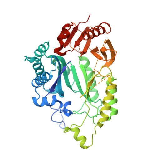
    The Elongator complex catalyzes posttranscriptional tRNA modifications by attaching carboxy-methyl (cm 5 ) moieties to uridine bases located in the wobble position. The catalytic subunit Elp3 is highly conserved and harbors two individual subdomains, a radical S-adenosyl methionine (rSAM) and a lysine acetyltransferase (KAT) domain. The details of its modification reaction cycle and particularly the substrate specificity of its KAT domain remain elusive. Here, we present the co-crystal structure of bacterial Elp3 (DmcElp3) bound to an acetyl-CoA analog and compare it to the structure of a monomeric archaeal Elp3 from Methanocaldococcus infernus (MinElp3). Furthermore, we identify crucial active site residues, confirm the importance of the extended N-terminus for substrate recognition and uncover the specific induction of acetyl-CoA hydrolysis by different tRNA species. In summary, our results establish the clinically relevant Elongator subunit as a non-canonical acetyltransferase and genuine tRNA modification enzyme.


  • Organizational Affiliation
    • Max Planck Laboratory, Malopolska Centre of Biotechnology (MCB), Jagiellonian University, Krakow, 30-387, Poland.

Macromolecule Content 

  • Total Structure Weight: 50.95 kDa 
  • Atom Count: 2,999 
  • Modeled Residue Count: 388 
  • Deposited Residue Count: 450 
  • Unique protein chains: 1

Macromolecules

Find similar proteins by:|  3D Structure
Entity ID: 1
MoleculeChains  Sequence LengthOrganismDetailsImage
ELP3 family, ELP3 family450Dehalococcoides mccartyi BTF08Mutation(s): 0 
Gene Names: btf_573
EC: 2.3.1.311
UniProt
Find proteins for A0A1C7D1B7 (Dehalococcoides mccartyi (strain CBDB1))
Explore A0A1C7D1B7 
Go to UniProtKB:  A0A1C7D1B7
Entity Groups
Sequence Clusters30% Identity50% Identity70% Identity90% Identity95% Identity100% Identity
UniProt GroupA0A1C7D1B7
Sequence Annotations
Expand
Reference Sequence

Experimental Data & Validation

Experimental Data

  • Method: X-RAY DIFFRACTION
  • Resolution: 2.70 Å
  • R-Value Free:  0.269 (Depositor), 0.272 (DCC) 
  • R-Value Work:  0.223 (Depositor), 0.231 (DCC) 
  • R-Value Observed: 0.225 (Depositor) 
Space Group: C 2 2 21
Unit Cell:
Length ( Å )Angle ( ˚ )
a = 73.54α = 90
b = 160.74β = 90
c = 92.99γ = 90
Software Package:
Software NamePurpose
PHENIXrefinement
XDSdata reduction
XSCALEdata scaling
PHENIXphasing

Structure Validation

View Full Validation Report


Ligand Structure Quality Assessment 


Entry History 

& Funding Information

Deposition Data


Funding OrganizationLocationGrant Number
PolandHoming/2016-2/14
PolandUMO-2015/19/B/NZ1/00343

Revision History  (Full details and data files)

  • Version 1.0: 2019-02-20
    Type: Initial release
  • Version 1.1: 2024-01-24
    Changes: Data collection, Database references, Refinement description