6I9Q | pdb_00006i9q

Structure of the mouse CD98 heavy chain ectodomain


Experimental Data Snapshot

  • Method: X-RAY DIFFRACTION
  • Resolution: 2.10 Å
  • R-Value Free: 
    0.218 (Depositor), 0.229 (DCC) 
  • R-Value Work: 
    0.175 (Depositor), 0.187 (DCC) 
  • R-Value Observed: 
    0.177 (Depositor) 

Starting Model: experimental
View more details

wwPDB Validation 3D Report Full Report

Validation slider image for 6I9Q

This is version 1.2 of the entry. See complete history

Literature

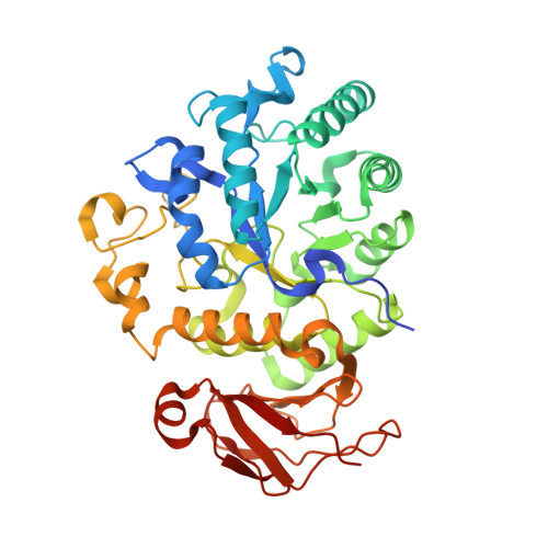
Structural differences between the ectodomains of murine and human CD98hc.

Deuschle, F.C.Schiefner, A.Skerra, A.

(2019) Proteins 87: 693-698

  • DOI: https://doi.org/10.1002/prot.25686
  • Primary Citation Related Structures: 
    6I9Q

  • PubMed Abstract: 

    The CD98 heavy chain (CD98hc) constitutes both a promising cell surface target for the treatment of cancers and a transcytosis receptor potentially useful for the brain delivery of therapeutics. However, pharmacokinetic studies and safety assessment of cognate antibodies or nonimmunoglobulin binding proteins in rodents is hampered by cross-species variability of both amino acid sequence and glycosylation pattern. Here, we report the crystal structure of the murine CD98hc extracellular domain and a comprehensive comparison with its human ortholog, revealing only one conserved surface patch that is neither shielded by glycosylation nor by the cell membrane with an accessible surface area typical for an antibody epitope. Our results imply the necessity of a surrogate approach for CD98hc-specific binding proteins with predictive power for clinical investigations.


  • Organizational Affiliation
    • Lehrstuhl für Biologische Chemie, Technische Universität München, Freising, Germany.

Macromolecule Content 

  • Total Structure Weight: 48.53 kDa 
  • Atom Count: 3,545 
  • Modeled Residue Count: 422 
  • Deposited Residue Count: 434 
  • Unique protein chains: 1

Macromolecules

Find similar proteins by:|  3D Structure
Entity ID: 1
MoleculeChains  Sequence LengthOrganismDetailsImage
4F2 cell-surface antigen heavy chain434Mus musculusMutation(s): 0 
Gene Names: Slc3a2Mdu1
UniProt
Find proteins for P10852 (Mus musculus)
Explore P10852 
Go to UniProtKB:  P10852
Entity Groups
Sequence Clusters30% Identity50% Identity70% Identity90% Identity95% Identity100% Identity
UniProt GroupP10852
Sequence Annotations
Expand
Reference Sequence

Experimental Data & Validation

Experimental Data

  • Method: X-RAY DIFFRACTION
  • Resolution: 2.10 Å
  • R-Value Free:  0.218 (Depositor), 0.229 (DCC) 
  • R-Value Work:  0.175 (Depositor), 0.187 (DCC) 
  • R-Value Observed: 0.177 (Depositor) 
Space Group: I 4 2 2
Unit Cell:
Length ( Å )Angle ( ˚ )
a = 97.384α = 90
b = 97.384β = 90
c = 218.98γ = 90
Software Package:
Software NamePurpose
XSCALEdata scaling
REFMACrefinement
PDB_EXTRACTdata extraction
XDSdata reduction
PHASERphasing

Structure Validation

View Full Validation Report


Entry History 

Deposition Data

Revision History  (Full details and data files)

  • Version 1.0: 2019-04-17
    Type: Initial release
  • Version 1.1: 2019-07-10
    Changes: Data collection, Database references
  • Version 1.2: 2024-01-24
    Changes: Data collection, Database references, Refinement description