Exposing the distinctive modular behavior of beta-strands and alpha-helices in folded proteins.
Wang, H., Logan, D.T., Danielsson, J., Oliveberg, M.(2020) Proc Natl Acad Sci U S A 117: 28775-28783
- PubMed: 33148805 Search on PubMedSearch on PubMed Central
- DOI: https://doi.org/10.1073/pnas.1920455117
- Primary Citation Related Structures:
6I69, 6I6E, 6I6I, 6I6O, 6I6S, 6I6U, 6I6W, 6I6Y - PubMed Abstract:
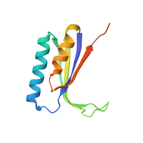
Although folded proteins are commonly depicted as simplistic combinations of β-strands and α-helices, the actual properties and functions of these secondary-structure elements in their native contexts are just partly understood. The principal reason is that the behavior of individual β- and α-elements is obscured by the global folding cooperativity. In this study, we have circumvented this problem by designing frustrated variants of the mixed α/β-protein S6, which allow the structural behavior of individual β-strands and α-helices to be targeted selectively by stopped-flow kinetics, X-ray crystallography, and solution-state NMR. Essentially, our approach is based on provoking intramolecular "domain swap." The results show that the α- and β-elements have quite different characteristics: The swaps of β-strands proceed via global unfolding, whereas the α-helices are free to swap locally in the native basin. Moreover, the α-helices tend to hybridize and to promote protein association by gliding over to neighboring molecules. This difference in structural behavior follows directly from hydrogen-bonding restrictions and suggests that the protein secondary structure defines not only tertiary geometry, but also maintains control in function and structural evolution. Finally, our alternative approach to protein folding and native-state dynamics presents a generally applicable strategy for in silico design of protein models that are computationally testable in the microsecond-millisecond regime.
- Department of Biochemistry and Biophysics, Arrhenius Laboratories of Natural Sciences, Stockholm University, S-106 91 Stockholm, Sweden.
Organizational Affiliation: