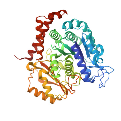
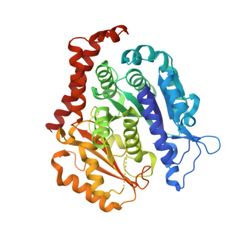
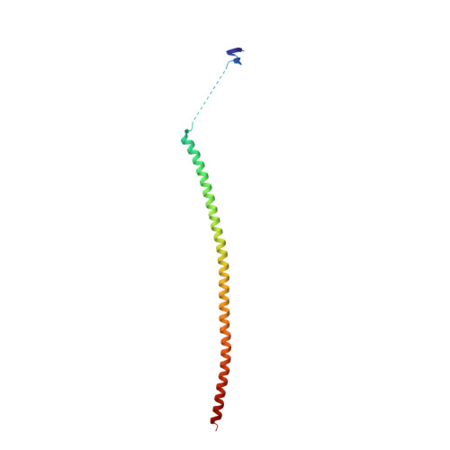
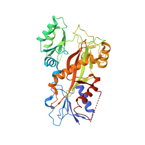
6I5C

Long wavelength native-SAD phasing of Tubulin-Stathmin-TTL complex


Experimental Data Snapshot

  • Method: X-RAY DIFFRACTION
  • Resolution: 2.95 Å
  • R-Value Free: 0.208 
  • R-Value Work: 0.170 
  • R-Value Observed: 0.172 

wwPDB Validation   3D Report Full Report


Ligand Structure Quality Assessment 


This is version 2.1 of the entry. See complete history


Literature

Long-wavelength native-SAD phasing: opportunities and challenges.

Basu, S.Olieric, V.Leonarski, F.Matsugaki, N.Kawano, Y.Takashi, T.Huang, C.Y.Yamada, Y.Vera, L.Olieric, N.Basquin, J.Wojdyla, J.A.Bunk, O.Diederichs, K.Yamamoto, M.Wang, M.

(2019) IUCrJ 6: 373-386

  • DOI: https://doi.org/10.1107/S2052252519002756
  • Primary Citation of Related Structures:  
    6I59, 6I5C

  • PubMed Abstract: 

    Native single-wavelength anomalous dispersion (SAD) is an attractive experimental phasing technique as it exploits weak anomalous signals from intrinsic light scatterers ( Z < 20). The anomalous signal of sulfur in particular, is enhanced at long wavelengths, however the absorption of diffracted X-rays owing to the crystal, the sample support and air affects the recorded intensities. Thereby, the optimal measurable anomalous signals primarily depend on the counterplay of the absorption and the anomalous scattering factor at a given X-ray wavelength. Here, the benefit of using a wavelength of 2.7 over 1.9 Å is demonstrated for native-SAD phasing on a 266 kDa multiprotein-ligand tubulin complex (T 2 R-TTL) and is applied in the structure determination of an 86 kDa helicase Sen1 protein at beamline BL-1A of the KEK Photon Factory, Japan. Furthermore, X-ray absorption at long wavelengths was controlled by shaping a lysozyme crystal into spheres of defined thicknesses using a deep-UV laser, and a systematic comparison between wavelengths of 2.7 and 3.3 Å is reported for native SAD. The potential of laser-shaping technology and other challenges for an optimized native-SAD experiment at wavelengths >3 Å are discussed.


  • Organizational Affiliation

    Swiss Light Source, Paul Scherrer Institut, Villigen PSI, 5232, Switzerland.


Macromolecules
Find similar proteins by:  (by identity cutoff)  |  3D Structure
Entity ID: 1
MoleculeChains Sequence LengthOrganismDetailsImage
Tubulin alpha-1B chain
A, C
440Bos taurusMutation(s): 0 
UniProt
Find proteins for P81947 (Bos taurus)
Explore P81947 
Go to UniProtKB:  P81947
Entity Groups  
Sequence Clusters30% Identity50% Identity70% Identity90% Identity95% Identity100% Identity
UniProt GroupP81947
Sequence Annotations
Expand
  • Reference Sequence
Find similar proteins by:  (by identity cutoff)  |  3D Structure
Entity ID: 2
MoleculeChains Sequence LengthOrganismDetailsImage
Tubulin beta-2B chain
B, D
431Bos taurusMutation(s): 0 
Gene Names: TUBB2B
UniProt
Find proteins for Q6B856 (Bos taurus)
Explore Q6B856 
Go to UniProtKB:  Q6B856
Entity Groups  
Sequence Clusters30% Identity50% Identity70% Identity90% Identity95% Identity100% Identity
UniProt GroupQ6B856
Sequence Annotations
Expand
  • Reference Sequence
Find similar proteins by:  (by identity cutoff)  |  3D Structure
Entity ID: 3
MoleculeChains Sequence LengthOrganismDetailsImage
Stathmin-4136Rattus norvegicusMutation(s): 0 
Gene Names: Stmn4
UniProt
Find proteins for P63043 (Rattus norvegicus)
Explore P63043 
Go to UniProtKB:  P63043
Entity Groups  
Sequence Clusters30% Identity50% Identity70% Identity90% Identity95% Identity100% Identity
UniProt GroupP63043
Sequence Annotations
Expand
  • Reference Sequence
Find similar proteins by:  (by identity cutoff)  |  3D Structure
Entity ID: 4
MoleculeChains Sequence LengthOrganismDetailsImage
TUBULIN-TYROSINE LIGASE379Gallus gallusMutation(s): 0 
Gene Names: TTL
Entity Groups  
Sequence Clusters30% Identity50% Identity70% Identity90% Identity95% Identity100% Identity
Sequence Annotations
Expand
  • Reference Sequence
Small Molecules
Ligands 7 Unique
IDChains Name / Formula / InChI Key2D Diagram3D Interactions
GTP
Query on GTP

Download Ideal Coordinates CCD File 
G [auth A],
P [auth C]
GUANOSINE-5'-TRIPHOSPHATE
C10 H16 N5 O14 P3
XKMLYUALXHKNFT-UUOKFMHZSA-N
ACP
Query on ACP

Download Ideal Coordinates CCD File 
W [auth F]PHOSPHOMETHYLPHOSPHONIC ACID ADENYLATE ESTER
C11 H18 N5 O12 P3
UFZTZBNSLXELAL-IOSLPCCCSA-N
GDP
Query on GDP

Download Ideal Coordinates CCD File 
K [auth B],
T [auth D]
GUANOSINE-5'-DIPHOSPHATE
C10 H15 N5 O11 P2
QGWNDRXFNXRZMB-UUOKFMHZSA-N
MES
Query on MES

Download Ideal Coordinates CCD File 
M [auth B],
N [auth B]
2-(N-MORPHOLINO)-ETHANESULFONIC ACID
C6 H13 N O4 S
SXGZJKUKBWWHRA-UHFFFAOYSA-N
CA
Query on CA

Download Ideal Coordinates CCD File 
I [auth A],
L [auth B],
O [auth B],
R [auth C],
U [auth D]
CALCIUM ION
Ca
BHPQYMZQTOCNFJ-UHFFFAOYSA-N
CL
Query on CL

Download Ideal Coordinates CCD File 
J [auth A],
S [auth C]
CHLORIDE ION
Cl
VEXZGXHMUGYJMC-UHFFFAOYSA-M
MG
Query on MG

Download Ideal Coordinates CCD File 
H [auth A],
Q [auth C],
V [auth F]
MAGNESIUM ION
Mg
JLVVSXFLKOJNIY-UHFFFAOYSA-N
Experimental Data & Validation

Experimental Data

  • Method: X-RAY DIFFRACTION
  • Resolution: 2.95 Å
  • R-Value Free: 0.208 
  • R-Value Work: 0.170 
  • R-Value Observed: 0.172 
  • Space Group: P 21 21 21
Unit Cell:
Length ( Å )Angle ( ˚ )
a = 104.24α = 90
b = 156.83β = 90
c = 179.54γ = 90
Software Package:
Software NamePurpose
PHENIXrefinement
PDB_EXTRACTdata extraction
XDSdata reduction
XSCALEdata scaling
SHELXCDphasing
BUCCANEERmodel building

Structure Validation

View Full Validation Report


Ligand Structure Quality Assessment 


Entry History 

Deposition Data

Revision History  (Full details and data files)

  • Version 1.0: 2019-03-13
    Type: Initial release
  • Version 1.1: 2019-03-20
    Changes: Data collection, Database references, Structure summary
  • Version 1.2: 2019-05-08
    Changes: Data collection, Database references
  • Version 1.3: 2019-05-29
    Changes: Data collection, Database references
  • Version 2.0: 2019-08-21
    Changes: Advisory, Atomic model, Data collection, Database references, Derived calculations, Polymer sequence, Source and taxonomy, Structure summary
  • Version 2.1: 2020-09-23
    Changes: Database references, Derived calculations