6I19 | pdb_00006i19

Crystal structure of Chlamydomonas reinhardtii thioredoxin h1


Experimental Data Snapshot

  • Method: X-RAY DIFFRACTION
  • Resolution: 1.38 Å
  • R-Value Free: 
    0.216 (Depositor), 0.217 (DCC) 
  • R-Value Work: 
    0.181 (Depositor), 0.182 (DCC) 
  • R-Value Observed: 
    0.183 (Depositor) 

Starting Model: experimental
View more details

wwPDB Validation 3D Report Full Report

Validation slider image for 6I19

This is version 1.2 of the entry. See complete history

Literature

Crystal Structure of Chloroplastic Thioredoxin f2 fromChlamydomonas reinhardtiiReveals Distinct Surface Properties.

Lemaire, S.D.Tedesco, D.Crozet, P.Michelet, L.Fermani, S.Zaffagnini, M.Henri, J.

(2018) Antioxidants (Basel) 7

  • DOI: https://doi.org/10.3390/antiox7120171
  • Primary Citation Related Structures: 
    6I19, 6I1C

  • PubMed Abstract: 

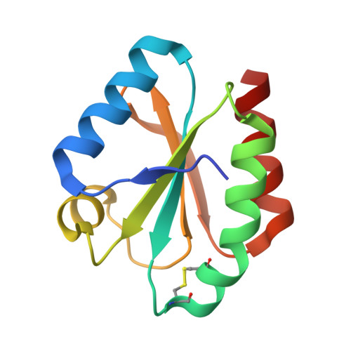
    Protein disulfide reduction by thioredoxins (TRXs) controls the conformation of enzyme active sites and their multimeric complex formation. TRXs are small oxidoreductases that are broadly conserved in all living organisms. In photosynthetic eukaryotes, TRXs form a large multigenic family, and they have been classified in different types: f, m, x, y, and z types are chloroplastic, while o and h types are located in mitochondria and cytosol. In the model unicellular alga Chlamydomonas reinhardtii , the TRX family contains seven types, with f- and h-types represented by two isozymes. Type-f TRXs interact specifically with targets in the chloroplast, controlling photosynthetic carbon fixation by the Calvin⁻Benson cycle. We solved the crystal structures of TRX f2 and TRX h1 from C. reinhardtii . The systematic comparison of their atomic features revealed a specific conserved electropositive crown around the active site of TRX f, complementary to the electronegative surface of their targets. We postulate that this surface provides specificity to each type of TRX.


  • Organizational Affiliation
    • Laboratoire de Biologie Moléculaire et Cellulaire des Eucaryotes, Institut de Biologie Physico-Chimique, Unité Mixte de Recherche 8226 CNRS Sorbonne Université, 13 rue Pierre et Marie Curie, 75005 Paris, France. lemaire@ibpc.fr.

Macromolecule Content 

  • Total Structure Weight: 23.72 kDa 
  • Atom Count: 2,033 
  • Modeled Residue Count: 222 
  • Deposited Residue Count: 226 
  • Unique protein chains: 1

Macromolecules

Find similar proteins by:|  3D Structure
Entity ID: 1
MoleculeChains  Sequence LengthOrganismDetailsImage
Thioredoxin H-type
A, B
113Chlamydomonas reinhardtiiMutation(s): 0 
Gene Names: TRXH
UniProt
Find proteins for P80028 (Chlamydomonas reinhardtii)
Explore P80028 
Go to UniProtKB:  P80028
Entity Groups
Sequence Clusters30% Identity50% Identity70% Identity90% Identity95% Identity100% Identity
UniProt GroupP80028
Sequence Annotations
Expand
Reference Sequence

Experimental Data & Validation

Experimental Data

  • Method: X-RAY DIFFRACTION
  • Resolution: 1.38 Å
  • R-Value Free:  0.216 (Depositor), 0.217 (DCC) 
  • R-Value Work:  0.181 (Depositor), 0.182 (DCC) 
  • R-Value Observed: 0.183 (Depositor) 
Space Group: P 31 2 1
Unit Cell:
Length ( Å )Angle ( ˚ )
a = 48.76α = 90
b = 48.76β = 90
c = 143.97γ = 120
Software Package:
Software NamePurpose
PHENIXrefinement
XDSdata reduction
XDSdata scaling
PHASERphasing

Structure Validation

View Full Validation Report


Entry History 

Deposition Data

Revision History  (Full details and data files)

  • Version 1.0: 2018-12-05
    Type: Initial release
  • Version 1.1: 2024-01-24
    Changes: Data collection, Database references, Refinement description
  • Version 1.2: 2024-10-09
    Changes: Structure summary