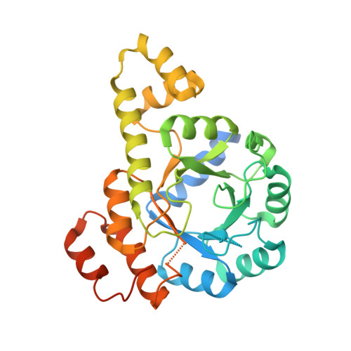
Crystal structure of the pseudoenzyme PDX1.2 in complex with its cognate enzyme PDX1.3: a total eclipse.
Robinson, G.C., Kaufmann, M., Roux, C., Martinez-Font, J., Hothorn, M., Thore, S., Fitzpatrick, T.B.(2019) Acta Crystallogr D Struct Biol 75: 400-415
- PubMed: 30988257 Search on PubMed
- DOI: https://doi.org/10.1107/S2059798319002912
- Primary Citation Related Structures:
6HX3, 6HXG, 6HYE - PubMed Abstract:
Pseudoenzymes have burst into the limelight recently as they provide another dimension to regulation of cellular protein activity. In the eudicot plant lineage, the pseudoenzyme PDX1.2 and its cognate enzyme PDX1.3 interact to regulate vitamin B 6 biosynthesis. This partnership is important for plant fitness during environmental stress, in particular heat stress. PDX1.2 increases the catalytic activity of PDX1.3, with an overall increase in vitamin B 6 biosynthesis. However, the mechanism by which this is achieved is not known. In this study, the Arabidopsis thaliana PDX1.2-PDX1.3 complex was crystallized in the absence and presence of ligands, and attempts were made to solve the X-ray structures. Three PDX1.2-PDX1.3 complex structures are presented: the PDX1.2-PDX1.3 complex as isolated, PDX1.2-PDX1.3-intermediate (in the presence of substrates) and a catalytically inactive complex, PDX1.2-PDX1.3-K97A. Data were also collected from a crystal of a selenomethionine-substituted complex, PDX1.2-PDX1.3-SeMet. In all cases the protein complexes assemble as dodecamers, similar to the recently reported individual PDX1.3 homomer. Intriguingly, the crystals of the protein complex are statistically disordered owing to the high degree of structural similarity of the individual PDX1 proteins, such that the resulting configuration is a composite of both proteins. Despite the differential methionine content, selenomethionine substitution of the PDX1.2-PDX1.3 complex did not resolve the problem. Furthermore, a comparison of the catalytically competent complex with a noncatalytic complex did not facilitate the resolution of the individual proteins. Interestingly, another catalytic lysine in PDX1.3 (Lys165) that pivots between the two active sites in PDX1 (P1 and P2), and the corresponding glutamine (Gln169) in PDX1.2, point towards P1, which is distinctive to the initial priming for catalytic action. This state was previously only observed upon trapping PDX1.3 in a catalytically operational state, as Lys165 points towards P2 in the resting state. Overall, the study shows that the integration of PDX1.2 into a heteromeric dodecamer assembly with PDX1.3 does not cause a major structural deviation from the overall architecture of the homomeric complex. Nonetheless, the structure of the PDX1.2-PDX1.3 complex highlights enhanced flexibility in key catalytic regions for the initial steps of vitamin B 6 biosynthesis. This report highlights what may be an intrinsic limitation of X-ray crystallography in the structural investigation of pseudoenzymes.
- Department of Botany and Plant Biology, University of Geneva, 30 Quai E. Ansermet, 1211 Geneva, Switzerland.
Organizational Affiliation: