


The crystal structure of type II Dehydroquinase from Butyrivibrio crossotus DSM 2876
Lapthorn, A.J., Ner, L., Roszak, A.W.To be published.
Experimental Data Snapshot
Starting Model: experimental
View more details
wwPDB Validation 3D Report Full Report

Entity ID: 1 | |||||
|---|---|---|---|---|---|
| Molecule | Chains | Sequence Length | Organism | Details | Image |
| 3-dehydroquinate dehydratase | 145 | Eshraghiella crossota DSM 2876 | Mutation(s): 0 Gene Names: aroQ, BUTYVIB_01550 EC: 4.2.1.10 | 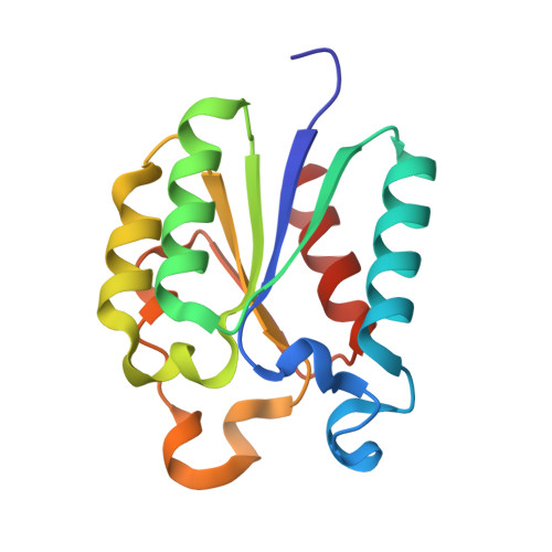 | |
UniProt | |||||
Entity Groups | |||||
| Sequence Clusters | 30% Identity50% Identity70% Identity90% Identity95% Identity100% Identity | ||||
| UniProt Group | D4S0D1 | ||||
Sequence AnnotationsExpand | |||||
Reference Sequence | |||||
| Ligands 5 Unique | |||||
|---|---|---|---|---|---|
| ID | Chains | Name / Formula / InChI Key | 2D Diagram | 3D Interactions | |
| ETE Download:Ideal Coordinates CCD File | B [auth A] | 2-{2-[2-2-(METHOXY-ETHOXY)-ETHOXY]-ETHOXY}-ETHANOL C9 H20 O5 ZNYRFEPBTVGZDN-UHFFFAOYSA-N |  | ||
| FLC Download:Ideal Coordinates CCD File | F [auth A] | CITRATE ANION C6 H5 O7 KRKNYBCHXYNGOX-UHFFFAOYSA-K |  | ||
| SER Download:Ideal Coordinates CCD File | C [auth A] | SERINE C3 H7 N O3 MTCFGRXMJLQNBG-REOHCLBHSA-N |  | ||
| SO4 Download:Ideal Coordinates CCD File | D [auth A], E [auth A] | SULFATE ION O4 S QAOWNCQODCNURD-UHFFFAOYSA-L |  | ||
| GOL Download:Ideal Coordinates CCD File | G [auth A] | GLYCEROL C3 H8 O3 PEDCQBHIVMGVHV-UHFFFAOYSA-N |  | ||
| Length ( Å ) | Angle ( ˚ ) |
|---|---|
| a = 79.476 | α = 90 |
| b = 79.476 | β = 90 |
| c = 72.033 | γ = 120 |
| Software Name | Purpose |
|---|---|
| REFMAC | refinement |
| PDB_EXTRACT | data extraction |
| XDS | data reduction |
| Aimless | data scaling |
| PHASER | phasing |
| Funding Organization | Location | Grant Number |
|---|---|---|
| Engineering and Physical Sciences Research Council | United Kingdom | EP/P00086X/1 |


